What appears to be a simple task for a cell—splitting in two—is indeed a complex series of engineering puzzles. To produce two working cells, a splitting cell has to maneuver its interiors in such a way that the appropriate components will end up in each cell.

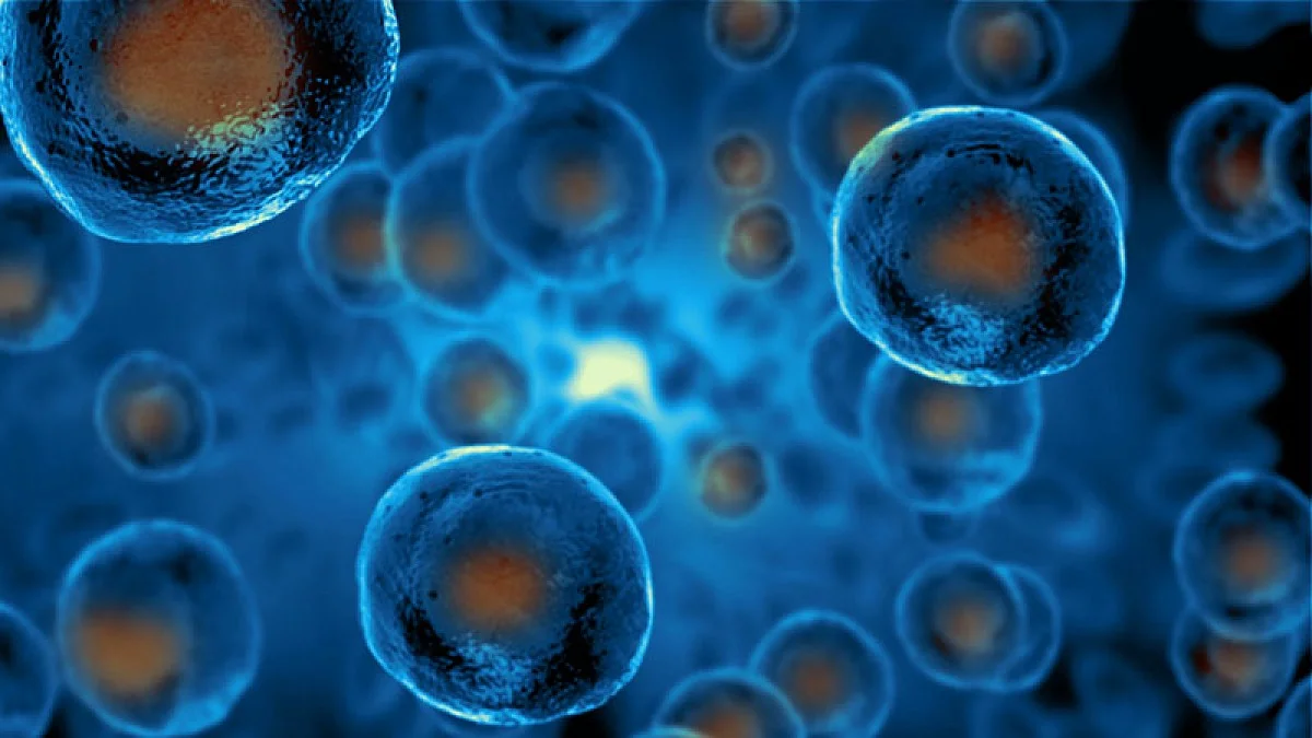
Illustration of stem cells. Image credit: University of Oregon
Ken Prehoda, a biochemist from the University of Oregon, is attempting to solve one of those basic puzzles: how a splitting stem cell slices out its membrane during the division process.
In new research, he and postdoctoral researcher Bryce LaFoya disclose how ready-to-divide stem cells produce a pool of extra membrane, accommodating the increased surface area needed for two cells. The pair explain their results in a paper published on April 27th, 2023 in Developmental Cell.
Animal cells are encircled by a thin membrane that creates a protective barrier surrounding the cell. Just before an animal cell splits, it becomes rounder, stated Prehoda, who is part of the College of Arts and Sciences.
Geometrically, a sphere is optimal for minimizing the amount of membrane. But to divide the cell in two, it gets squeezed and the surface area sharply increases.”
Ken Prehoda, Biochemistry, University of Oregon
In this regard, cell division seems to be pinching a balloon, only for the fact that balloons can stretch as they alter the shape. The membrane of the cell is not stretchy like the membrane of a balloon, but it still requires to be able to expand in some way to accommodate squeezing jointly and pinching off a new cell.
The research team of Prehoda concentrated on this challenge in neural stem cells, which are the cells that produce the cells in the nervous system. As they continue to churn out new cells, stem cells split asymmetrically: The stem cell retains a majority of the cell’s material, giving only a little bit away to the new sister cell.
Prehoda and LaFoya employed a spinning disk confocal microscope fitted with advanced super-resolution technology to look into the brains of developing fruit flies. LaFoya observed that neural stem cell membranes were decorated with small protrusions and folds, “kind of like the extra skin on wrinkly dog breeds like shar-pei and bulldogs,” he states.
LaFoya recognized that the neural stem cell membrane utilizes these “wrinkles” to store up the membrane for when it is required during division. As LaFoya observed the division of the cells, he was astonished to notice the wrinkles gather on one side prior to division. The placing of the wrinkles found where the new cell could form and where the cell could grow.
Although dividing unevenly enables the neural stem cell to give up just a fraction of its resources, membrane partitioning is made a particularly complicated problem by the asymmetry. LaFoya highlighted that the cell has got an outstanding solution to that engineering puzzle’s part.
“Making the extra membrane beforehand and positioning it properly before division is an elegant solution that we hadn’t envisioned before starting this project,” he states.
That is only one of several questions Prehoda and his lab are attempting to answer using cutting-edge microscopy techniques, enabling them to observe cells in new levels of detail. Owing to this new technology, Prehoda stated, “We have an embarrassment of riches, and lots of new imagery to explore.”
Source:
Journal reference:
LaFoya, B., & Prehoda, K. E. (2023). Consumption of a polarized membrane reservoir drives asymmetric membrane expansion during the unequal divisions of neural stem cells. Developmental Cell. doi.org/10.1016/j.devcel.2023.04.006.