Reviewed by Danielle Ellis, B.Sc.Jun 14 2023
Lung mesenchymal cells, which are crucial components of the lung’s distinctive structure, also play a significant role in disease and injury recovery, but research into their biology and how they start disorders like pulmonary fibrosis is limited.

Image Credit: Boston University Chobanian & Avedisian School of Medicine.
While experimental models have assisted in identifying certain regulators of lung mesenchyme behavior, it is unclear how lung mesenchyme is recognized during human development.
To fully understand these mechanisms, scientists have created an in-vitro induced pluripotent stem cell (iPSC)-based model system for the generation and study of early lung-specific mesenchyme, which has the potential to aid in understanding basic mechanisms governing tissue-specific mesenchymal fate decisions as well as future applications in regenerative medicine.
Our study has implications for the study of lung diseases, such as pulmonary fibrosis and interstitial lung diseases that arise from dysfunction of the part of the lung known as mesenchyme. These diseases currently have very limited treatment options and we hope our model system will provide new tools to understand what goes wrong in these diseases and to screen for better drugs.”
Darrell Kotton MD, Study Corresponding Author and David C. Seldin Professor, Medicine, Boston University School of Medicine
Darrell Kotton is also the Director of the BU/Boston Medical Center for Regenerative Medicine (CReM).

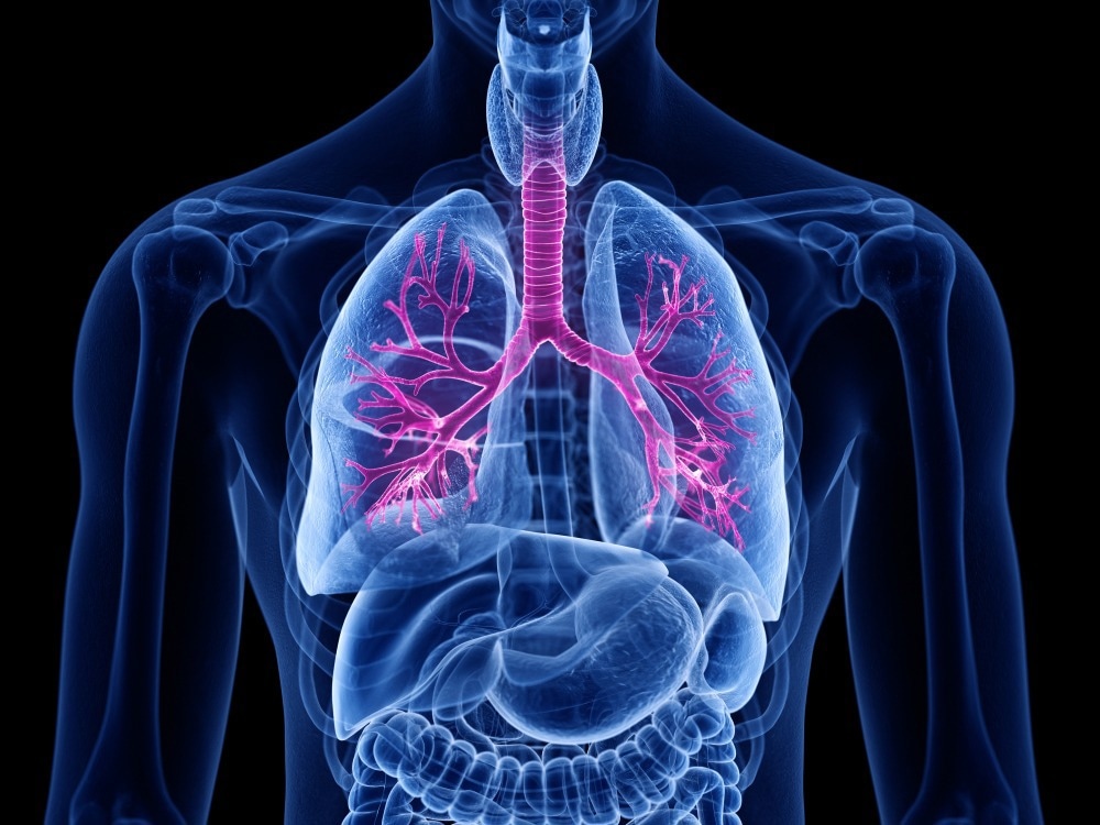
Image Credit: SciePro/Shutterstock.com
The researchers employed an iPSC line containing a lung mesenchyme-specific fluorescent reporter, which meant that cells that became lung mesenchymal fluoresced green. Using this model, scientists examined numerous growth factors and small compounds for their ability to trigger pathways known to be involved in lung development.
They discovered that boosting the retinoic acid and hedgehog signaling pathways, both of which are known to be important in embryonic development, resulted in the highest percentage of green fluorescent cells, indicating the presence of lung mesenchyme.
Researchers then separated those cells and contrasted their gene expression profile to that of primary cells from the experimental model’s embryonic lungs to see how comparable these cells are to primary lung mesenchymal cells. Finally, they employed their recombinant organoid system to see if these cells might act as lung mesenchyme.
An important role of the developing lung mesenchyme in the experimental model is their ability to interact with and signal to the neighboring epithelium. We found that our engineered cells can recapitulate some of those signaling interactions, suggesting that they have functional capacity.”
Andrea Alber PhD, Study First Author and Postdoctoral Fellow, Boston University School of Medicine
The part of the study in which the engineered lung mesenchymal cells are coupled with lung epithelial cells in culture dishes (recombinants) is particularly exciting, according to the researchers, because it results in organoids, living cells assembled together in a 3D culture gel that enables researchers to understand how cells are organized and communicate.
We are now working to apply these types of new organoid models to better understand pulmonary fibrosis.”
Andrea Alber PhD, Study First Author and Postdoctoral Fellow, Boston University School of Medicine
Source:
Journal reference:
Alber, A. B., et al. (2023). Directed differentiation of mouse pluripotent stem cells into functional lung-specific mesenchyme. Nature Communications. doi.org/10.1038/s41467-023-39099-9.