Reviewed by Danielle Ellis, B.Sc.Jul 31 2023
Neurons are the cells that constitute brain circuits and employ chemicals and electricity to receive and transmit information that allows the body to perform anything from thinking to sensing to movement. An axon is a long fiber in neurons that delivers information to neighboring neurons.
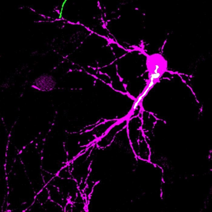
 The image shows a spiny stellate neuron (magenta) and its Golgi apparatus (white) in the mouse barrel cortex at postnatal day 5. The study found that the Golgi apparatus shows a biased distribution in one side of the neuron only during the postnatal critical period of circuit reorganization. This lateral Golgi polarity instructs the direction in which the neuron expands its dendrites for establishing precise neuronal circuit. Image Credit: NAKAGAWA, Naoki & IWASATO, Takuji, National Institute of Genetics, ROIS
Dendrites are branch-like structures that extend out from the cell body and receive information from axons.
Early postnatal brain development includes dendritic refinement, in which dendrites are shaped to connect with the proper axons in a certain way.
In a study that was just published, researchers provide evidence demonstrating how a mechanism within rodent neurons involving the Golgi apparatus initiates dendritic refinement with the aid of neuronal activity received by a receptor of a neurotransmitter known as N-methyl-D-aspartate-type glutamate receptor (NMDAR).
Cell Reports published the study on July 28th, 2023.
We wanted to find the cellular mechanism that underlies activity-dependent neuronal circuit reorganization during early postnatal brain development. The potential contribution of the dynamics of subcellular structures, such as organelles, to this process has been largely overlooked.”
Naoki Nakagawa, Assistant Professor, National Institute of Genetics
The Golgi apparatus, which serves as a focal point for material movement inside the cell, is the organelle in question. By controlling the direction of intracellular transport, the Golgi apparatus’ location within a cell creates cell polarity.
Nakagawa added, “Golgi-mediated cell polarity has been known to play an important role in earlier embryonic developmental events, such as the mitosis, migration, and differentiation of cells. However, during the postnatal critical period of circuit reorganization, we did not know if the Golgi polarity is changed by neuronal activity or if the Golgi polarity change contributes to the remodeling of neuronal circuits.”

Image Credit: Giovanni Cancemi/Shutterstock.com
Spiny stellate neurons from rodents were examined by researchers better to understand the function of Golgi polarity in dendritic refinement. These neurons are located in the barrel cortex, a region of the rodent brain that interprets tactile data from whiskers. Asymmetrical dendrites on spiny stellate neurons in the barrel cortex face the center of the barrel.
During the first week following birth, this particular dendritic structure is developed via refining depending on neural activity. Spiny stellate neurons were used to produce the Golgi-targeted fluorescent protein to monitor the location of the Golgi apparatus during postnatal development.
The Golgi apparatus in spiny stellate cells were apically polarized in the first three days after birth, but by day five following birth, it had become laterally polarized toward the barrel center. The lateral polarization decreased by day 15, when the asymmetric dendritic pattern was already well-established.
The location of the Golgi apparatus within the dendrites was another area of investigation. Dendrites with Golgi apparatus at the base or within were longer and more branched than those without.
NMDAR signals are necessary for lateral Golgi polarization, which guides the appropriate dendritic refinement. These were evident when the NMDAR signal or the Golgi polarity was disrupted. The form and orientation of the dendrites changed in both situations in an unexpected way.
Future work will focus on learning more about how the Golgi apparatus impacts neuron development.
Nakagawa concluded, “We want to know how the neuronal activity moves the Golgi apparatus to the proper positions within neurons and how the Golgi polarization enables proper patterning of neuronal dendrites. We think that addressing these questions will provide a better understanding of what is happening within neurons during postnatal development and how it promotes circuit reorganization, which is an essential step for constructing sophisticated neuronal circuits in the brain.”