Reviewed by Danielle Ellis, B.Sc.Aug 16 2023
Biomolecular condensates are loosely organized assemblies that carry out a variety of vital tasks inside all live cells. However, the mechanism by which proteins and other biomolecules combine to create these assemblies within cells is not fully known.
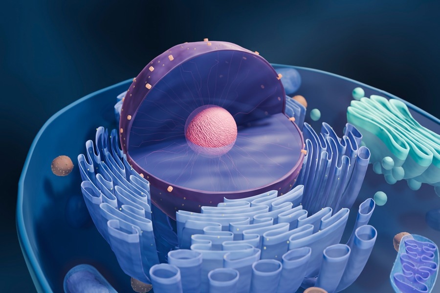
 MIT biologists discovered that a scaffolding protein called TCOF1 is responsible for the formation of a biomolecular condensate called the fibrillar center, which forms within the cell nucleolus, represented in pink inside the purple nucleus. Image Credit: iStock
Now, Massachusetts Institute of Technology (MIT) researchers have shown that one of these condensates, which develops inside a cell organelle called the nucleolus, is formed by a single scaffolding protein. This condensate cannot form without the protein TCOF1.
The discoveries could shed light on a significant evolutionary change in the nucleolus’ structure that occurred about 300 million years ago. The nucleolus, which aids in the construction of ribosomes, was formerly separated into two divisions.
The nucleolus in amniotes, which are mammals, birds, and reptiles, evolved a condensate that serves as a third compartment. Biologists still do not completely comprehend the reasons for this change.
If you look across the tree of life, the basic structure and function of the ribosome has remained quite static; however, the process of making it keeps evolving. Our hypothesis for why this process keeps evolving is that it might make it easier to assemble ribosomes by compartmentalizing the different biochemical reactions.”
Eliezer Calo, Study Senior Author and Associate Professor, Massachusetts Institute of Technology
It might be simpler for researchers to examine the fibrillar center’s activity in cells now that they are aware of how it originates. The researchers claim that the results also provide light on how other condensates may have first developed in cells.
The study’s lead authors are former MIT graduate students Nima Jaberi-Lashkari PhD ‘23 and Byron Lee PhD ‘23. Cell Reports published the study on August 15th, 2023. The study also includes Fardin Aryan, a former research associate at MIT.
Condensate Information
Membrane-bound organelles like lysosomes and mitochondria carry out many essential cellular operations, while membraneless condensates also carry out crucial jobs including gene control and stress response. These condensates can form when needed and then dissipate after completing their purpose.
Calo added, “Almost every cellular process that is essential for the functioning of the cell has been associated somehow with condensate formation and activity. However, it is not very well sorted out how these condensates form.”
In a study published in 2022, Calo and his coworkers discovered a protein area that seems to be involved in the formation of condensates. In that study, low-complexity regions (LCRs), or segments of proteins from many different species, were found and compared using computational methods. LCRs are repeating sequences of a single amino acid with a few additional amino acids thrown in.
That research also demonstrated the presence of many glutamate-rich LCRs in the nucleolar protein TCOF1, which can serve as a scaffold for biomolecular assemblies.
In this recent study, the researchers discovered that condensates occur whenever TCOF1 is expressed in cells. These condensates invariably contain the proteins that are typically present in the fibrillar center (FC) of the nucleolus, a distinct condensate. It is known that the FC participates in the synthesis of ribosomal RNA, an essential part of ribosomes, the cell complex in charge of producing all cellular proteins.
The fibrillar center, which is crucial for the construction of ribosomes, did not exist in single-celled creatures, invertebrates, or the first vertebrates (fish), but it only started to emerge some 300 million years ago.
According to the latest study, TCOF1 was necessary for this change from a “bipartite” to a “tripartite” nucleolus. Without TCOF1, the researchers discovered that cells only create two nucleolar compartments. Additionally, the addition of TCOF1 to zebrafish embryos, which typically have bipartite nucleoli, allowed the researchers to stimulate the creation of a third compartment.
“More than just creating that condensate, TCOF1 reorganized the nucleolus to acquire tripartite properties, which indicates that whatever chemistry that condensate was bringing to the nucleolus was enough to change the composition of the organelle,” Calo noted.
Scaffold Evolution
The glutamate-rich low-complexity sections of TCOF1 are the crucial part of the protein that aids in the formation of scaffolds, according to the researchers. These LCRs appear to interact with other glutamate-rich areas of nearby TCOF1 molecules, facilitating the assembly of the proteins into a scaffold that can subsequently draw more proteins and biomolecules to create the fibrillar center.
What’s really exciting about this work is that it gives us a molecular handle to control a condensate, introduce it into a species that doesn’t have it, and also get rid of it in a species that does have it. That could really help us unlock the structure-to-function relationship and figure out what is the role of the third compartment.”
Nima Jaberi-Lashkari PhD, Former Graduate Student, Massachusetts Institute of Technology
According to the results of this study, the scientists have a theory that cellular condensates that first appeared earlier in evolutionary history may have been initially supported by a single protein, similar to how TCOF1 supports the fibrillar center, but eventually developed to become more complicated.
Calo further stated, “Our hypothesis, which is supported by the data in the paper, is that these condensates might originate from one scaffold protein that behaves as a single component, and over time, they become multicomponent.”
ALS, Huntington’s disease, and cancer have all been related to the development of particular kinds of biomolecular condensates.
In all of these situations, what our work poses is this question of why are these assemblies forming, and what is the scaffold in these assemblies? And if we can better understand that, then I think we have a better handle on how we could treat these diseases.”
Byron Lee, Former Graduate Student, Massachusetts Institute of Technology
Source:
Journal reference:
Jaberi-Lashkari, N., et al. (2023). An evolutionarily nascent architecture underlying the formation and emergence of biomolecular condensates. Cell Reports. doi.org/10.1016/j.celrep.2023