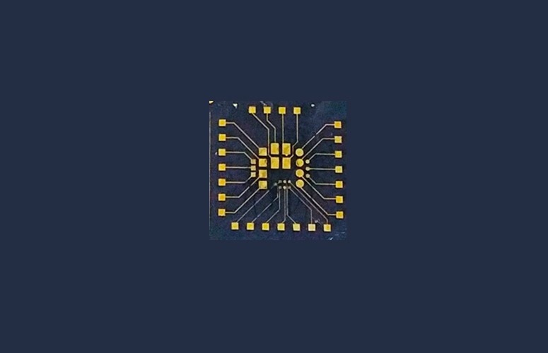
In the book titled “Microfluidic Systems for Cancer Diagnosis,” the UPV/EHU’s Microfluidics Cluster group outlines a method for constructing a bioelectronic device. This device features gold electrodes coated with an intelligent polymer that possesses the capability to non-invasively capture and release cells in a controlled manner, all while monitoring these processes through conventional electrical measurements.
 Microfabricated bioelectronic device for cell capture and release in which 24 gold microelectrodes of different sizes can be seen. Device size 20 × 20 mm. Image Credit: Microfludics Cluster UPV/EHU
These initial stages mark the initial strides toward the creation of versatile platforms for early cancer screening.
Metastasis stands as the primary cause of fatality in cancer, a process wherein a cell departs from the primary tumor enters the bloodstream and lymphatic system, and eventually arrives at distant organs.
The non-invasive retrieval of these circulating tumor cells is of paramount importance for various aspects of cancer research, including the study of cell biology, cancer diagnosis, prognosis, and drug development. Typically, cancer cells within the bloodstream are present at notably low concentrations compared to other cell types, and the conventional approaches for their viable collection are labor-intensive.
We wanted to come up with a device capable of concentrating cancer cells in order to detect their concentration.”
Janire Sáez, Ikerbasque Research Professor, Microfluidics Cluster Group, University of the Basque Country
To date, the biosensors designed for this task, which are instruments for measuring biological or chemical parameters containing a biological component, have had the drawback of causing harm to cells during the capture and release procedures.
In response, the Microfluidics Cluster group has innovatively merged smart materials with the realm of bioelectronics, which involves the application of carbon-based semiconductors. This integration enables the monitoring of the capture and release of cancer cells without inflicting damage.
The methodology has been extensively elucidated within a chapter featured in the book titled “Microfluidic Systems for Cancer Diagnosis.” This book is oriented toward the scientific community and delves into the most recent advancements in microfluidic technologies tailored for cancer diagnosis and monitoring.
Serving as a valuable resource, the book serves as an ideal reference for constructing microfluidic devices within laboratory settings, particularly those devised for cancer diagnostics. Furthermore, it actively fosters the advancement of novel and enhanced diagnostic instruments.
We show a bioelectronic device consisting of microfabricated gold electrodes coated with a smart polymer (which reacts to temperature changes) and which enables the non-invasive capture and release of circulating tumor cells to be made, and the simultaneous electrical and optical monitoring of the entire process to be conducted”.
Janire Sáez, Ikerbasque Research Professor, Microfluidics Cluster Group, University of the Basque Country
First Steps Towards a Major Breakthrough
“Our tests were performed in culture media; we did not use actual patient samples, but commercial cells sustained in cell culture. We confirmed that our device enabled us to capture and release the cells,” detailed the researcher.
The scientists are working to tailor the polymer so as to suit various cell types. The device “is the outcome of collaboration with a group at the University of Cambridge, with whom we continue to collaborate, and where the device is currently being applied to samples from esophageal cancer patients. Through this device, cancer cells are being selectively reconcentrated in order to detect their concentration,” adds Sáez.
The first steps towards developing platforms for cancer screening. This could be a good step forward because they generally involve low-cost technologies and can be mass-produced. The idea is to use this type of technology for early cancer diagnosis.”
Janire Sáez, Ikerbasque Research Professor, Microfluidics Cluster Group, University of the Basque Country
The Microfluidics Cluster group is concentrating its research on the development of “micrometric structures for bioelectronic devices of this type. We are also developing 3D systems to create 'organ-on-a-chip' systems (biomimetic systems that simulate organs in the human body),” she concludes.
Source:
Journal reference:
Saez, J., et al. (2023). Capture and Release of Cancer Cells Through Smart Bioelectronics. Methods in Molecular Biology. doi.org/10.1007/978-1-0716-3271-0_21.