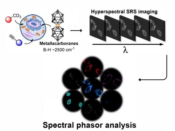
Researchers from CÚRAM at the University of Galway, in collaboration with their counterparts at the Centre for Molecular Nanometrology at the University of Strathclyde, have released findings that reveal the intricate mechanisms within cells.
 High resolution visualization of cell components using three chemical probes. Image Credit: CÚRAM/Pau Farras
Recently published in the German scientific journal Angewandte Chemie, this research delves into a more comprehensive understanding of the intricate interconnections among components within cells. This topic has long been a focal point for scientists worldwide, providing valuable insights into the behaviors of certain diseases.
The team, employing SRS microscopy for cellular visualization, has successfully tackled the challenge of obtaining clear images of individual processes.
Previous attempts at multiplex optical detection in live cells were hindered by time-consuming analyses and difficulties arising from poor image quality. Moreover, these efforts were limited in tracking processes, often requiring physical alterations to the cell for clearer images.
In contrast, the presented work utilizes dyes that necessitate no adjustments to the cell itself, ensuring completion within minutes. Remarkably, it allows simultaneous tracking of up to nine different aspects of cell structure. This represents a significant breakthrough, surpassing the limitations of previous work, which could only track seven processes.
This work will provide scientists with a tool to garner lots of information out of cells within a short space of time. This has the potential to assist in understanding how current drugs developed for a range of applications are fighting disease and even provide hints on how to improve treatments.”
Dr. Pau Farras, Study Lead Author and Associate Professor, Inorganic Chemistry, School of Biological and Chemical Sciences, University of Galway
Farras is also the Principal Investigator at CÚRAM SFI Research Center for Medical Devices.
Source:
Journal reference:
Kueckmann, T., et al. (2023) Expanding the Range of Bioorthogonal Tags for Multiplex Stimulated Raman Scattering Microscopy. Angewandte Chemie. doi.org/10.1002/anie.202311530.