A collaborative team from EPFL and Harvard has introduced an advanced artificial intelligence (AI) technique to track neurons within animals in motion. The new CNN-based AI hopes to address current challenges in decoding the brain activity of flexible organisms like worms.
 Two-dimensional projection of 3D volumetric brain activity recordings in C. elegans. (VIDEO) Video Credit: Mahsa Barzegar-Keshteli (EPFL)
The study, spearheaded by Sahand Jamal Rahi at EPFL’s School of Basic Sciences, has been published in Nature Methods.
Neural Imaging and AI
Historically, whole-brain imaging with single-cell resolution has been pivotal in studying neural circuits in various organisms. However, the dynamic nature of these studies, particularly in animals performing natural behaviors, poses a challenge.
The brain's movement and deformation within flexible organisms make the segmentation and tracking of individual neurons complex. To address this, the team set out to automate this process, overcoming the limitations of manual annotation, which, though reliable, is considerably time-consuming.
Neuron Segmentation and Tracking
The team's AI-based method centers around a convolutional neural network (CNN), a form of AI known for excelling at pattern recognition in images. The CNN was trained to recognize and comprehend image patterns through convolution, analyzing small picture segments for features like edges, colors, or shapes.
The notable mention of this technology is the introduction of 'targeted augmentation', a method that synthesizes artificial annotations from a limited number of manual annotations. This technique allows the CNN to learn the brain's internal deformations in different postures, thereby significantly reducing the need for manual annotation.

Image Credit: Hussmann/Shutterstock.com
Targettrack: A Comprehensive Pipeline for Neural Analysis
An end-to-end pipeline called 'Targettrack' was developed, which learns the brain-wide deformations in each experiment and adapts to the visual appearance of each animal and the specific imaging conditions. By using Targettrack, a substantial increase in throughput can be achieved compared to full manual annotation, reducing the time needed for manual annotations and proofreading by 50-fold for challenging brain imaging problems.
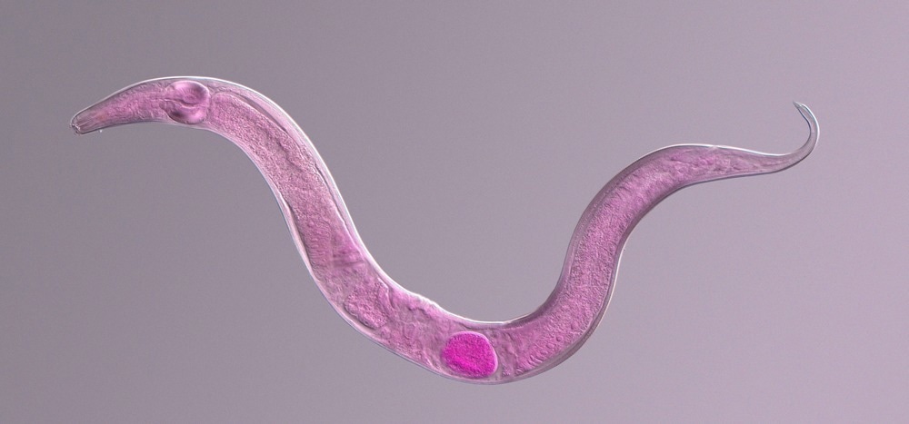
Tested on the nematode Caenorhabditis elegans, known for its compact neural system and popularity as a model organism in neuroscience, the CNN demonstrated its effectiveness in identifying neurons as key points or 3D volumes.
Furthermore, researchers were able to observe intricate patterns in interneuron dynamics, including changes in neuronal response to varying stimuli like odor bursts. The method's versatility and efficiency make it a valuable tool not only for C. elegans imaging but also for tracking neurons in other moving and deforming entities.
The breakthrough has the potential to accelerate research in brain imaging and deepen our understanding of neural circuits and behaviors."
Sahand Jamal Rahi, School of Basic Sciences, EPFL
Targettrack is also designed with the user in mind, with an accessible graphical interface that integrates targeted augmentation and a streamlined workflow that lays the foundation for gaining in-depth insight into neural circuits and behaviors.
Source:
Journal reference:
Park, C.F., Barzegar-Keshteli, M., Korchagina, K. et al. Automated neuron tracking inside moving and deforming C. elegans using deep learning and targeted augmentation. Nat Methods (2023). https://doi.org/10.1038/s41592-023-02096-3