Reviewed by Danielle Ellis, B.Sc.Dec 11 2023
Scientists at Tokyo Metropolitan University have gauged the fluctuations in oxygenated hemoglobin levels in the brains of infants in response to tactile stimuli.
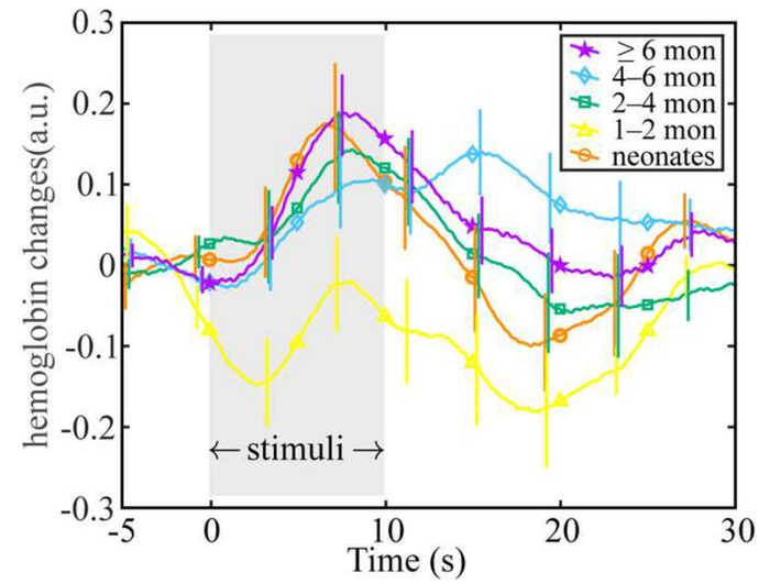
 The team found the same peaked response in oxygenated hemoglobin levels in infants of different ages over their first year. Image Credit: Tokyo Metropolitan University.
Utilizing spectroscopy techniques and external sensors positioned on the scalps of sleeping infants, they discovered that the timing of peak hemoglobin levels remains consistent across different infant ages, while the extent of variation over time differs. These findings contribute valuable insights into the physiological development of infants.
The initial stage of a newborn’s life is characterized by rapid and intricate developmental changes, a realm that neuroscience is unraveling through cutting-edge measurement techniques.
Functional magnetic resonance imaging (fMRI) and near-infrared spectroscopy (NIRS) of the brain are pivotal tools in this exploration. NIRS, involving an array of external sensors on an infant's head, enables scientists to monitor the changing flow of various compounds in the brain.
The research places particular emphasis on hemoglobin, the crucial oxygen-carrying component in blood. As the brain reacts to external stimuli such as light, heat, and touch, oxygen is dispatched to the brain. NIRS facilitates the distinction between levels of oxygenated and deoxygenated hemoglobin, providing insights into the brain's response.
Led by Assistant Professor Yutaka Fuchino, a team from Tokyo Metropolitan University applied NIRS to investigate how infant brains react to touch. Studying infants ranging from zero to one year old, the team affixed sensors to the infants’ scalps during sleep and observed how oxygenated hemoglobin levels changed as their limbs experienced a gentle shake.
Interestingly, infants of varying ages exhibited a similar response time, with a minor hemoglobin peak occurring a few minutes after the stimulus initiation. This uniformity in response across the first year implies that the factors influencing the speed of the response are present at birth.
However, the amplitude of the signal, representing the range of variation, differed significantly between infants of different age groups. This non-linear change, dipping for infants aged 1 to 2 months before increasing with age, could be attributed to various factors, including the initial rise in oxygen levels at birth, temporary anemia in the first months, and subsequent recovery.
The team’s work underscores how NIRS analysis of hemoglobin levels in the brain can serve as a reflection of developmental changes. Future studies are expected to delve into comparing other physiological responses to blood flow dynamics, offering deeper insights into the intricate development of the brain and its interactions with the body.
Source:
Journal reference:
Fuchino, Y., et al. (2023) Developmental changes in neonatal hemodynamics during tactile stimulation using whole-head functional near-infrared spectroscopy. NeuroImage. doi.org/10.1016/j.neuroimage.2023.120465.