A team of researchers from The University of Texas at Austin, led by SMU nanotechnology expert MinJun Kim, developed a less costly method to detect nuclease digestion, one of the essential processes in many nucleic acid sensing applications, such as those used to identify COVID-19.
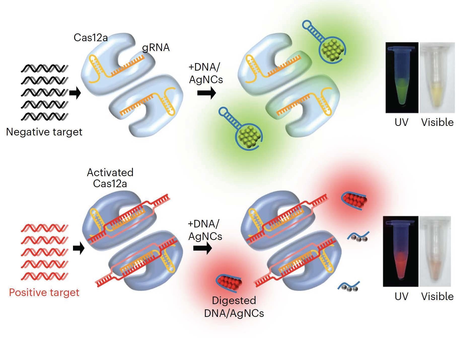
 Subak reporters, based on a special class of what are known as fluorescent silver nanoclusters, change color when they detect that so-called nuclease digestion has happened, which is one of the critical steps in the identification of disease-causing pathogens, such as COVID-19. These Subak reporters are much more cost-effective than the way nuclease activity is now detected. Image Credit: Tim Yeh/The University of Texas at Austin
The main technique for determining the pathogens responsible for infectious illnesses is nucleic acid detection. It is crucial to lower the cost of these tests since during the COVID-19 pandemic, millions of PCR tests were performed daily throughout the world.
According to a study that was published in the journal Nature Nanotechnology, this inexpensive device, known as Subak, is useful for determining when nuclease digestion—the process by which an enzyme called nuclease breaks down nucleic acids, such as DNA or RNA, into smaller fragments has taken place.
Producing a Fluorescence Resonance Energy Transfer (FRET) probe, the conventional method of determining nuclease activity, is 62 times more expensive than a Subak reporter.
Subak reporter is more cost-effective and simpler than FRET-based systems, offering an alternative method for detecting nuclease activity, and many nucleic acid detection methods today, such as PCR and DETECTR, still rely on the use of FRET probes in their final steps.”
MinJun Kim, Robert C. Womack Chair Professor, Department of Mechanical Engineering, Southern Methodist University
Kim is also a Principal Investigator of the BAST Lab.
The simpler assay, or test, DETECTR (DNA Endonuclease-Targeted CRISPR Trans Reporter), which uses CRISPR-Cas nuclease for pathogenic DNA detection, is in contrast to PCR. In the DETECTR assay, Kim and the UT Austin researchers have successfully substituted the Subak reporter for the FRET probe, significantly lowering the test's cost.
The foundation of Subak reporters is a unique class of fluorescent silver nanoclusters. They are composed of 13 silver atoms encircling a brief DNA strand. They are an organic/inorganic composite nanomaterial, ranging in size from 1 to 3 nm, that is too small to be seen with the unaided eye.
At this length scale, nanomaterials can display various hues and be very luminous, like quantum dots. Fluorescent nanoparticles have been used in biosensing devices like the Subak reporter and TV displays.
The Subak reporters were engineered by lead researcher Tim Yeh, an Associate Professor of Biomedical Engineering at the Cockrell School of Engineering at UT Austin, and his colleagues to release a distinct color when they are broken down by nucleases.
These DNA-templated silver nanoclusters initially emit green fluorescence, but undergo a remarkable color-switching to bright red when DNA is fragmented by nucleases, and the color change of Subak reporters is easily visible under a UV lamp, even though the actual device is miniscule.”
MinJun Kim, Robert C. Womack Chair Professor, Department of Mechanical Engineering, Southern Methodist University
The production of Subak reporters only costs $1 per nanomolecule. On the other hand, Kim stated that the production of a single nano molecule of FRET costs $62, requiring the use of many fluorescent dyes that are needed to get the desired results.
To develop and characterize the DNA/AgNC silver nanoclusters, Yeh collaborated with Kim and Madhav L. Ghimire, the Dean's Postdoctoral Fellow at SMU's Moody School of Graduate and Advanced Studies. This involved intensifying the red and green fluorescent colors both before and after nuclease fragmentation.
The process of characterization includes verifying the nanoclusters' dimensions, composition, and stability in particular settings.
Optimization of these low-cost detectors is essential to monitor their fluorescence properties, ensuring nanocluster’s stability, controlling size and structure, and most importantly to enhance their sensitivity and selectivity in various environmental conditions, making them more reliable for the sensing purpose.”
Madhav L. Ghimire, Postdoctoral Fellow, Moody School of Graduate and Advanced Studies, Southern Methodist University
The team intends to study whether the Subak reporter may be used as a probe for additional biological targets in addition to testing it further for nuclease digestion.