Scientists in Sweden were able to uncover the molecular mechanisms behind the development of lesions in multiple sclerosis by employing sophisticated techniques. The latest findings are detailed in the journal Cell by a team of researchers from Karolinska Institutet and Stockholm University.
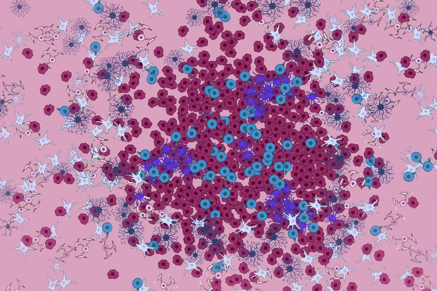
 The illustration shows the spatial distribution in lesions of the body's immune cells and different types of glial cells - oligodendrocytes, astrocytes, and microglia. Image Credit: Amagoia Agirre.
The global MS population is estimated to be about 1.85 million. The immune cells in the body target the glial cells, or so-called oligodendrocytes, which are the cells that makeup myelin in this condition.
Myelin prevents impulses from traveling between nerve cells as quickly, which can lead to symptoms including decreased feeling and disorientation. Brain and spinal cord lesions are characteristic of multiple sclerosis.
We wanted to understand which cells are part of the lesions and their dynamics over time.”
Petra Kukanja, Study Co-First Author, Department of Biochemistry and Biophysics, Karolinska Institutet
Kukanja is from the research group of Professor Gonçalo Castelo-Branco.
The researchers employed a technique known as in situ sequencing, created in Stockholm University's research group by Professor Mats Nilsson. This entails using the active genes in a certain cell to analyze and identify the cells that make up a tissue segment.
This pattern shows how various cell types are distributed inside the tissue, in this case, the spinal cord as well as how the cells communicate with one another. Samples were obtained at various stages from human MS patients and mice that were deliberately engineered to exhibit symptoms similar to MS to investigate the development of the lesions.
We analyzed simultaneously 239 genes and saw that active lesions in mice were built up centrifugally in two dimensions, with immune cells in the middle and different types of glial cells around them.”
Christoffer Mattsson Langseth, Study Co-First Author, Department of Biochemistry and Biophysics, Stockholm University
It was evident from the mice that the injuries started in the spinal cord and went all the way to the brain. 260 genes were examined simultaneously in spinal cord samples from four MS patients who passed away, allowing for the identification of the cellular structure of the lesions. The scientists also discovered new lesions and sub-structures inside the lesions.
Victims of Immune Cell Attacks
In the past, oligodendrocytes were primarily thought to be the targets of immune cell assaults.
The fact that they are active in the outer regions of lesions but also the entire brain and spinal cord raises the question of whether they dampen the disease or drive it.”
Petra Kukanja, Study Co-First Author, Department of Biochemistry and Biophysics, Karolinska Institutet
Because MS is such a diverse condition, the researchers hope to apply the same methodology to examine samples from many more people. What the lesions look like in patients with various therapies is another topic.
The two researchers draw attention to the broad and productive cooperation between their research groups, which has been going on since 2019. They have each contributed their area of expertise, with Karolinska Institutet contributing knowledge on the cell biology underlying MS and Stockholm University contributing cutting-edge in situ sequencing technology and novel approaches to complex image analysis.
Source:
Journal reference:
Kukanja, P., et al. (2024) Cellular architecture of evolving neuroinflammatory lesions and multiple sclerosis pathology. Cell. doi.org/10.1016/j.cell.2024.02.030