A group from IRB Barcelona and the University of Barcelona has created a novel bioinformatics instrument to recognize the chromosomal changes characteristic of cancerous cells. This new detection system, called QATS (QuAntification of Toroidal nuclei in biological imageS), is a computational biological imaging processing tool that can automatically identify and quantify the phenotypes linked to chromosomal instability in cancer cell nuclei, which will improve tumor research and classification.
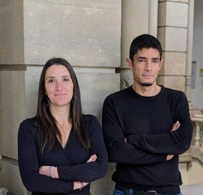
 Dr Mauvezin and Dr Pons. Image Credit: IRB Barcelona
The study, published in the journal Bioinformatics, is signed by Professor Dr. Caroline Mauvezin of the UB’s Faculty of Medicine and Health Sciences and IDIBAPS, as well as researcher Dr. Carles Pons of the Barcelona IRB.
Identifying Chromosomal Changes in Cancer Cells
Chromosomal instability is widespread in solid tumors and has been related to cancer’s start and development, as well as cancer cell metastasis. This process, produced by changes in the number and shape of chromosomes during cell division, can alter DNA and impact the entire cellular machinery.
Furthermore, chromosomal instability promotes tumor formation and development while also increasing intra-tumor heterogeneity and resistance to anti-tumor therapy.
Cancer cells can survive with a high degree of chromosomal instability. The new QATS tool is a predictive system for identifying and quantifying toroidal nuclei, which are novel indicators of chromosomal instability in biological imaging.
Toroidal nuclei are phenotypically different from normal nuclei, since these present a ring shape and a void with cytosolic material. In the field of research, these have been recently characterized as important biomarkers of chromosomal instability, and they represent an innovative pathway to understand and fight cancer. Traditionally, the level of chromosomal instability in cancer cells has only been assessed by quantifying micronuclei, which are irregular structures derived from the cell nucleus that may contain chromosomes or chromosomal fragments.”
Dr. Caroline Mauvezin, Professor, Faculty of Medicine and Health Sciences, University of Barcelona
Dr. Carles Pons, a member of the Structural Bioinformatics and Network Biology Laboratory at IRB Barcelona, added, “Therefore, integrating the strategy to assess toroidal nuclei into research and clinical practice has immense potential for tumor stratification and the design of patient-specific treatments.”
Currently, the QATS system has demonstrated its ability to recognize and quantify toroidal nuclei in preclinical cancer cell lines.
The study authors concluded, “In the future, the application of QATS in more complex biological scenarios — human tissue samples from patient biopsies — will represent a breakthrough for the scientific and medical communities to improve cancer diagnosis and patient treatment.”
Source:
Journal reference:
Pons, C., and Mauvezin, C. (2024). QATS: an ImageJ plugin for the quantification of toroidal nuclei in biological images. Bioinformatics. doi.org/10.1093/bioinformatics/btae026