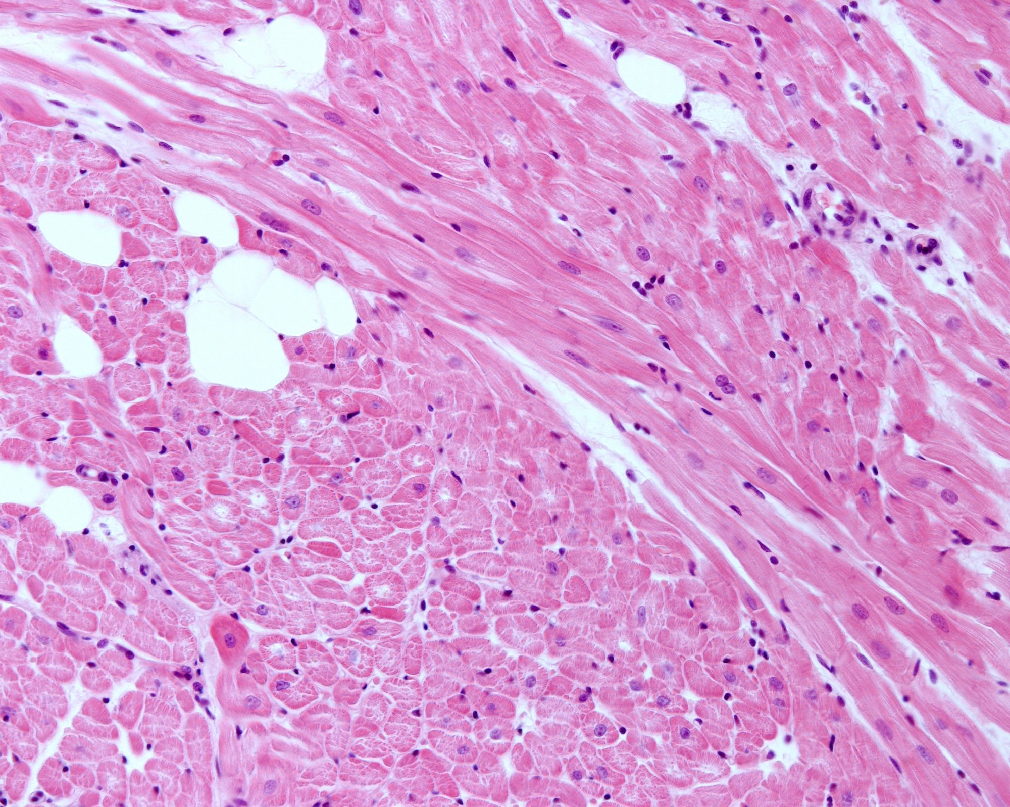
Fetal cardiomyocytes depend largely on glycolysis for energy, while adult cardiomyocytes receive all their energy through oxidative phosphorylation carried out in the mitochondria. The maturation of fetal and newborn cardiomyocytes involves a downregulation of glycolysis, an increase in mitochondrial biogenesis, and the loss of mitotic ability.
In a recent study published in The Journal of Clinical Investigation, researchers from the United States used murine models to investigate whether forcing adult cardiomyocytes to reverse from the oxidative phosphorylation phenotype to the glycolysis phenotype would also restore the mitotic ability in these cells.
 Study: Mitochondria regulate proliferation in adult cardiac myocytes. Image Credit: Jose Luis Calvo/Shutterstock.com
Background
The primary function of mitochondria is to produce energy in the form of adenosine triphosphate or ATP through the process of oxidative phosphorylation. However, mitochondria are also involved in various other biological processes, such as calcium signaling, redox signaling, and stress signal generation.
Some of the biomolecules required for histone modification and other epigenetic changes to the deoxyribonucleic acid (DNA), and the substrates for nucleotide, protein, and lipid synthesis are also produced by mitochondria.
Since the heart is one of the most metabolically active organs, cardiomyocytes are highly dependent on the mitochondria for ATP.
Fetal cardiomyocytes, however, obtain their energy primarily from glycolysis while retaining the ability to undergo mitosis. Maturation of infant cardiomyocytes occurs through a gradual downregulation of glycolysis, activation of mitochondrial biogenesis, and a loss of mitotic ability.
About the Study
In the present study, the researchers aimed to test whether the retention of the glycolytic phenotype was linked to the proliferative state and that the shift from glycolysis to oxidative phosphorylation in cardiomyocytes was responsible for the transition from the proliferative state to the mature, post-mitotic state.
The researchers used murine models, where they deleted the gene Uqcrfs1 (Ubiquinol-Cytochrome C Reductase, Rieske Iron-Sulfur Polypeptide 1), which codes for the Rieske iron-sulfur protein (RISP) essential for electron transfer.
Given that this gene is non-essential for cell survival but is related to mitochondrial function, the researchers hypothesized that the deletion of Uqcrfs1 would gradually transition the cell towards a glycolytic phenotype as the existing RISP proteins get utilized by the mitochondria.
The tamoxifen-inducible Cre recombinase system, which is used to observe the temporal and tissue-specific changes of gene deletion in murine models, was used to observe the effects of Uqcrfs1 knock-out.
The residual levels of RISP in the heart were measured at one, two, and two and a half months to determine if the protein levels were depleting.
Mitochondrial respiration was also measured from mitochondria isolated from cardiomyocytes harvested from the mice at 60 days.
The researchers also compared cultured neonatal cardiomyocytes from wild-type mice with the cardiomyocytes from the RISP knockout mice to determine the role of RISP in oxidative phosphorylation and the consequences of decreasing RISP levels on mitochondrial function.
Additionally, cardiac function was assessed through echocardiography and measurements of left ventricle dimensions and ejection fraction, posterior wall thickness, cardiac output, and heart rate. Blood pressure measurements were also taken non-invasively.
Cardiac tissue samples were also subjected to immunoblotting and histological examinations using various stains such as hematoxylin/eosin, periodic acid schiff, wheat germ agglutinin, and 4′,6-diamidino-2-phenylindole or DAPI.
Major Findings
The study found that with decreasing levels of RISP, mitochondrial function in cardiomyocytes decreased, and glucose utilization in the cells increased, indicating a gradual shift to the glycolytic phenotype.
Simultaneously, the cardiomyocytes underwent hyperplastic remodeling, where the cell number increased without accompanying cellular hypertrophy, indicating the activation of a proliferative state.
Furthermore, the energy supply in the cell was maintained, and the activation of the mammalian target of rapamycin (mTOR) indicated active cellular proliferation.
The absence of 5' adenosine monophosphate-activated protein kinase (AMPK) activation also suggested that ATP levels were adequate because AMPK is a sensor that gets activated in low ATP and mitochondrial stress conditions.
Ribonucleic acid (RNA) sequencing also indicated that the genes involved in cardiomyocyte proliferation and cardiac development were upregulated. The metabolomic analyses showed a decrease in alpha-ketoglutarate levels, an essential component of Kreb’s cycle involved in oxidative phosphorylation. An increase in methyltransferase activity also implied an increase in cell proliferation.
Furthermore, the hyperplastic remodeling that the cardiomyocytes underwent after transitioning from oxidative phosphorylation to glycolysis only involved cell proliferation, with no inflammation, fibrosis, or cellular hypertrophy.
Hearts that underwent induced ischemic injury showed migration of new cardiomyocytes to the site of the infarction after RISP depletion, suggesting the potential use of this method for cardiac regeneration.
Conclusions
To summarize, the findings showed that deletion of the gene coding for RISP gradually converted adult cardiomyocytes from an oxidative phosphorylation phenotype to a glycolytic one in mice.
Along with this change in the energy production process, the cell also underwent a shift into a proliferative state, resulting in an increase in cardiomyocyte number, but without the accompanying hypertrophy, inflammation, or fibrosis.
These results indicate that mitochondria are key to adult cardiomyocytes' proliferation regulation.