Scientists are working to create quicker, more quantitative, and more publicly available methods of seeing biomolecules in living cells to speed up biotechnology discoveries, such as the production of lifesaving drugs.
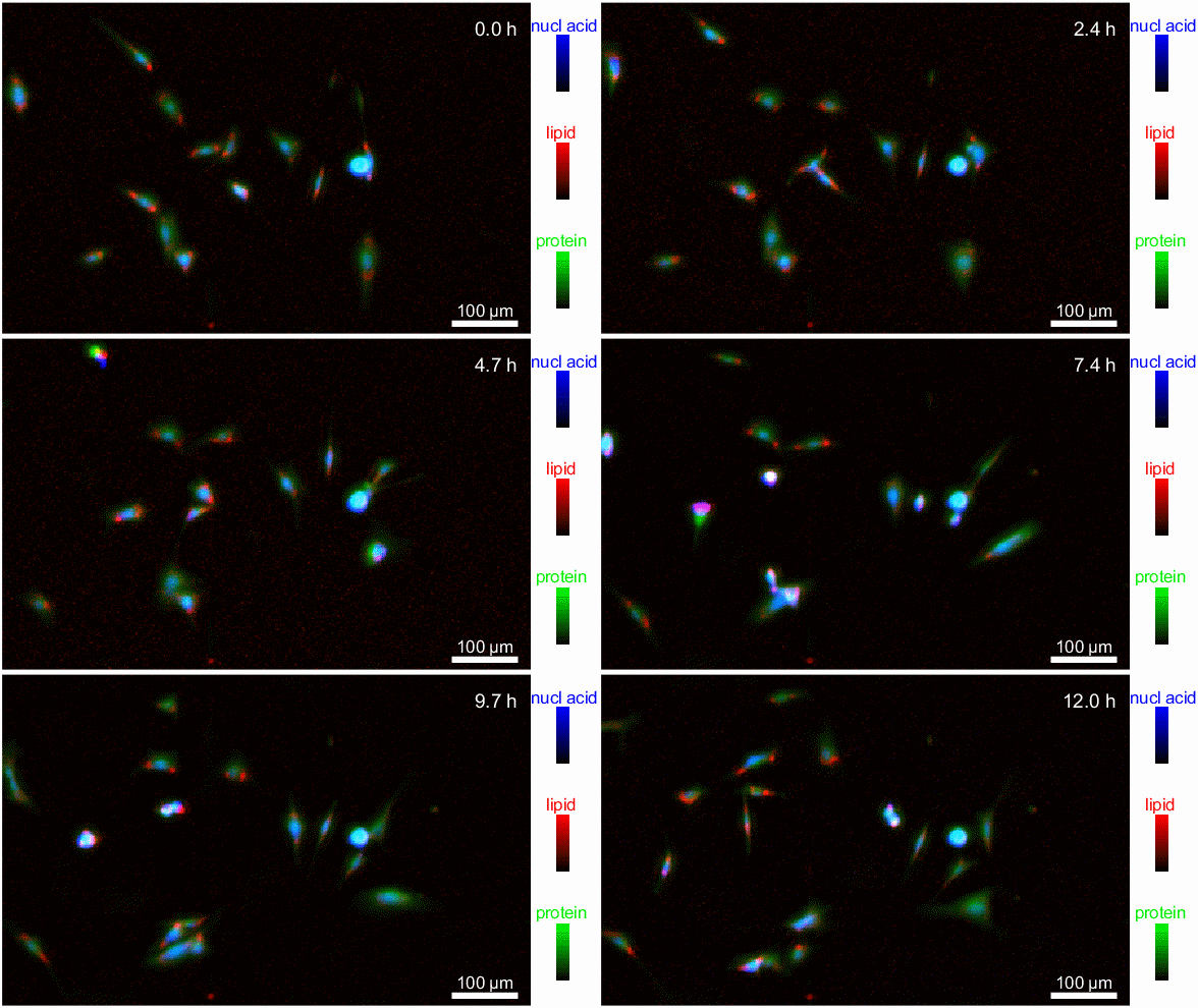
 An image of biomolecules, such as nucleic acids, lipids and proteins, in live cells using an imaging technique called infrared (IR) transmission microscopy. Image Credit: Y. Lee/NIST
Researchers at the National Institute of Standards and Technology (NIST) have developed a new method that uses infrared (IR) light to capture clear images of biomolecules inside cells. This was previously impossible due to water's tendency to absorb infrared radiation.
The novel technology eliminates the obscuring effects of water in IR-based measurements, allowing researchers to estimate the concentrations of essential biomolecules in cells, such as proteins that control cell activity. The capacity to monitor changes in living cells has the potential to accelerate progress in biomanufacturing, cell therapy research, drug development, and other fields.
Their findings were reported in Analytical Chemistry.
Infrared radiation is light that is slightly beyond the human eye's visual range. Although humans cannot see infrared light, we can detect it as heat. In IR microscopy, the material of interest absorbs light from a variety of wavelengths in the infrared spectrum.
Scientists examine and evaluate a sample's IR absorption spectrum, resulting in a collection of "fingerprints" for identifying molecules and other chemical structures. However, water, the most common molecule both within and outside cells, absorbs infrared extensively, masking the absorption of other biomolecules in cells.
To comprehend this optical masking effect, consider when an airplane passes above near the Sun. The Sun makes it difficult to view the airplane with the naked eye, but if a special Sun-blocking filter is applied, it can seen clearly in the sky.
In cancer cell therapy, for example, when cells from a patient’s immune system are modified to better recognize and kill cancer cells before being reintroduced back to the patient, one must ask, ‘Are these cells safe and effective?’ Our method can be helpful by providing additional insight with respect to biomolecular changes in the cells to assess cell health,”
Young Jong Lee, Chemist, National Institute of Standards and Technology
Lee invented a patented approach for compensating for IR water absorption with an optical element. The approach, known as solvent absorption compensation (SAC), was combined with a hand-built infrared laser microscope to view fibroblast cells, which help to build connective tissue.
Over a 12-hour observation period, researchers were able to identify groupings of biomolecules (proteins, lipids, and nucleic acids) at various stages of the cell cycle, including cell division. While this may appear to be a lengthy period, it is ultimately faster than existing methods, which need beam time at a huge synchrotron facility.
This new technology, known as SAC-IR, is label-free, which means it does not use any dyes or fluorescent markers that might injure cells and generate inconsistent findings across labs.
The SAC-IR approach allowed NIST researchers to determine the absolute mass of proteins, nucleic acids, lipids, and carbohydrates in a cell. The methodology might help provide the groundwork for standardized techniques of measuring biomolecules in cells, which could be important in biology, medicine, and industry.
Lee added, “In cancer cell therapy, for example, when cells from a patient’s immune system are modified to better recognize and kill cancer cells before being reintroduced back to the patient, one must ask, ‘Are these cells safe and effective?’ Our method can be helpful by providing additional insight with respect to biomolecular changes in the cells to assess cell health.”
Other possible applications include utilizing cells for drug screening, whether to identify novel medications or to assess the safety and efficacy of a therapeutic candidate. For example, this approach might aid in determining the efficacy of novel medications by measuring the absolute concentrations of various biomolecules in a large number of individual cells, as well as analyzing how different types of cells react to the drugs.
The researchers intend to improve the approach so that it can detect other important chemicals, such as DNA and RNA, more accurately. The approach might also assist offer specific answers to fundamental problems in cell biology, such as whether biomolecule fingerprints correspond to cell viability — that is, whether the cell is living, dying, or dead.
Lee concluded, “Some cells are preserved in a frozen state for months or years, then thawed for later use. We don’t yet fully understand how best to thaw the cells while maintaining maximum viability. With our new measurement capabilities, we may be able to develop better processes for cell freezing and thawing by looking at their infrared spectra.”
Source:
Journal reference:
Chang, Y., et al. (2024) Benchtop IR Imaging of Live Cells: Monitoring the Total Mass of Biomolecules in Single Cells. Analytical Chemistry. doi.org/10.1021/acs.analchem.4c02108