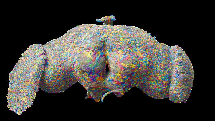
Study: Neuronal wiring diagram of an adult brain. Image Credit: Nature
Background
The evolution of brains around 500 million years ago played a crucial role in the development of complex behaviors. However, technological challenges made it difficult for scientists to map neuronal and synaptic connections within different brain regions.
With recent advances in electron microscopic brain imaging, starting in the 2000s, researchers began creating detailed wiring diagrams of animal brains.
Studies on Drosophila melanogaster (fruit flies) revealed that more than 100,000 neurons and nearly 100 million synapses control behaviors like vision, movement, and even social interactions.
These diagrams have improved our understanding of neural circuits and their relation to behaviors like navigation, sensory processing, and memory, opening a window into the functioning of the mammalian brain.
About the Study
In the present study, a large team of researchers, including The FlyWire Consortium, headed by scientists at Princeton University, used a combination of imaging and artificial intelligence (AI) tools to build the first whole-brain connectome or wiring diagram of a female fruit fly brain.
The FlyWire Consortium is a human-AI collaboration consisting of neurobiologists, proofreaders, and computer scientists who came together to reconstruct the entire connectome of a fruit fly brain.
The study involved electron microscopy imaging of one-week-old female fruit flies, after which neuron boundary-detecting networks and various algorithms were used to align the segments in the images to the cells.
To further improve the analysis, they incorporated previously acquired images of adult fruit fly brains to segment neuropils, which are synapse-rich regions in the brain.
The researchers then calculated each neuropil’s volume and classified these synapse-rich regions into different brain sections. Synapses were assigned to their respective neuropils based on location, and any synapses that could not be precisely matched were assigned to the nearest neuropil within a 10-micrometer range.
The neuron segmentation data underwent careful review and correction by the team of proofreaders to ensure accuracy. Machine learning tools were employed to predict synapse types and neurotransmitter identities.
The proofreading process focused primarily on central brain neurons with high synapse counts and visible nuclei. Neurons that exhibited significant changes during proofreading were flagged for additional review, ensuring meticulous correction.
Citizen scientists also played a key role in the study by helping to annotate and categorize neurons based on function and connectivity.
Further machine learning analysis was applied to predict the neurotransmitter type at each synapse, providing insights into the communication pathways within the fruit fly brain.
Major Findings
The study successfully mapped the entire neuronal wiring or connectome of a female fruit fly brain, providing neurobiologists with an advanced method to analyze the interconnectivity of neural pathways with specific regions of the brain, such as the suboesophageal zone and the optic lobes, which are important for processing sensory information and visual signals, respectively.
The female fruit fly connectome, consisting of 139,255 neurons and 54.5 million synapses, provided crucial insights into neuronal connections and communication pathways and was a significant achievement in comparison with previous brain mapping efforts involving Caenorhabditis elegans and Drosophila larvae.
The project also identified 8,400 distinct cell types within the connectome and revealed how neurons connect to the ventral nerve cord, which plays a critical role in controlling movement. This neural network map is accessible via The FlyWire Consortium’s connectome data explorer.
The connectome has also facilitated simulations of neural functions, such as visual and taste processing. The researchers believe that these simulations will improve as new data on fly neuron biophysics, such as inhibitory synapse properties, become available.
Data acquisition through electron microscopy and advancements in computational tools have allowed for improved image alignment and reconstruction, and FlyWire’s automated segmentation and extensive proofreading helped achieve a high accuracy of close to 99.2%.
However, the study also discussed the limitations in the detection of small synaptic connections, which the researchers believe could be addressed with future technological advancements.
Conclusions
Overall, through a comprehensive map of the neural networks of the female fruit fly brain, the researchers highlighted the potential applications of a complete connectome in advancing our understanding of brain function and communication between different regions of the brain.
The insights into the flow of information and processing of sensory signals provided by the connectome are a significant contribution to deciphering mammalian brains.