Human bodies are exceptional at adapting to shifting situations. For example, thanks to a process known as homeostasis, the internal temperature remains constant at 37 °C during the summer and the winter. This concealed balancing act is critical for survival, allowing animals to maintain consistent internal conditions even as the exterior world changes.
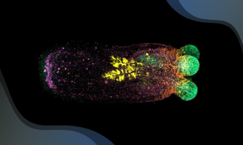
 By using four different colors to label specific genes, scientists can visualise where each gene is active in the sea anemone’s body, helping them understand the body organisation in both intact and regenerating animals. Image Credit: Matthew Benton/EMBL
However, recent research from the Ikmi Group at EMBL Heidelberg demonstrates that homeostasis can go beyond internal regulation to actively remodel an organism’s structure.
The starlet sea anemone (Nematostella vectensis) has exceptional regenerating potential. If its head or foot is cut off, it sprouts another one. Cut it in half, and each half becomes a fully functional anemone.
While some regeneration creatures, such as salamanders and fish, work to restore damaged parts proportionately to what remains, this sea anemone takes a different strategy. It reshapes its entire body to keep the same general shape, even if this requires altering areas that were not harmed. This characteristic is also found in flatworms and other animals that can regenerate their entire body.
Regeneration is about restoring function after tissue loss or damage. Most research studies mainly consider patterns and sizes in regeneration, but our findings show that maintaining shape is also crucial—and it is something the organism actively controls.”
Aissam Ikmi, Study Senior Author and Group Leader, European Molecular Biology Laboratory (EMBL)
Stephanie Cheung, a doctoral researcher in Ikmi’s lab, observed something unexpected. When a sea anemone’s foot was injured, Cheung discovered cell division at the wound site and unanticipated cell division at the other end of the body—the mouth. This demonstrated that the anemone was transmitting messages throughout its body in reaction to the injury.
To study this, the researchers combined spatial transcriptomics with sophisticated imaging. This enabled them to determine which genes were active in various areas of the anemone's body during regeneration. What they discovered was surprising: the injury caused molecular alterations near and distant from the wound. Cells moved and tissues reorganized, essentially changing the entire organism.
The level of body reshaping varied according to the severity of the damage. Losing a foot generated minor modifications, whereas cutting the anemone in half resulted in major remodeling. The researchers discovered a family of enzymes known as metalloproteases, which became more active as more tissue was destroyed. These enzymes were active at the wound site and throughout the body, assisting in tissue realignment.
Metalloprotease activity has never been shown before in animals like this. I had to design and optimize experimental conditions for Nematostella based on the sparse literature available from other species. This took some time, but the final results were very rewarding.”
Petrus Steenbergen, Study Lead Author and Senior Research Technician, European Molecular Biology Laboratory (EMBL)
The breakthrough occurred when the researchers realized these alterations were intended to restore the anemone's original shape. They discovered that the anemone's aspect ratio, or length-to-width ratio, had recovered to its pre-injury proportions. So, even if the anemone shrank due to an injury, it retained its shape.
Ikmi added, “We were able to witness the body-wide coordination that drives this remodeling. This proportional response allows the anemone to restore its shape, highlighting how organisms like Nematostella interpret and respond to tissue loss in a way that’s scaled to the damage incurred.”
This research was a team effort. Rik Korswagen’s team at the Hubrecht Institute in the Netherlands contributed to implementing spatial transcriptomics in sea anemones. Oliver Stegle’s team at EMBL Heidelberg and the German Cancer Research Center (DKFZ) provided bioinformatics expertise and the statistical approaches required to cope with spatial gene expression data.
It was a pleasure to puzzle out the findings of the study together by uniting the team’s expertise in data analysis and cell biology. This work was a truly collaborative journey, and I am glad I was part of it.
Tobias Gerber, Study Lead Author and Postdoctoral Fellow, European Molecular Biology Laboratory (EMBL)
Looking ahead, Ikmi and his team are eager to explore new questions.
Ikmi stated, “The next big question is why maintaining shape is so important. And how does the organism sense its own shape? How does it know what it currently looks like?”
They are excited to study the magnificent starlet sea anemone to learn more about how organisms heal and maintain equilibrium.
Source:
Journal reference:
Cheung, S., et. al. (2024) Systemic coordination of whole-body tissue remodeling during local regeneration in sea anemones. Developmental Cell. doi.org/10.1016/j.devcel.2024.11.001