.jpg) By Dr. Chinta SidharthanReviewed by Lexie CornerDec 13 2024
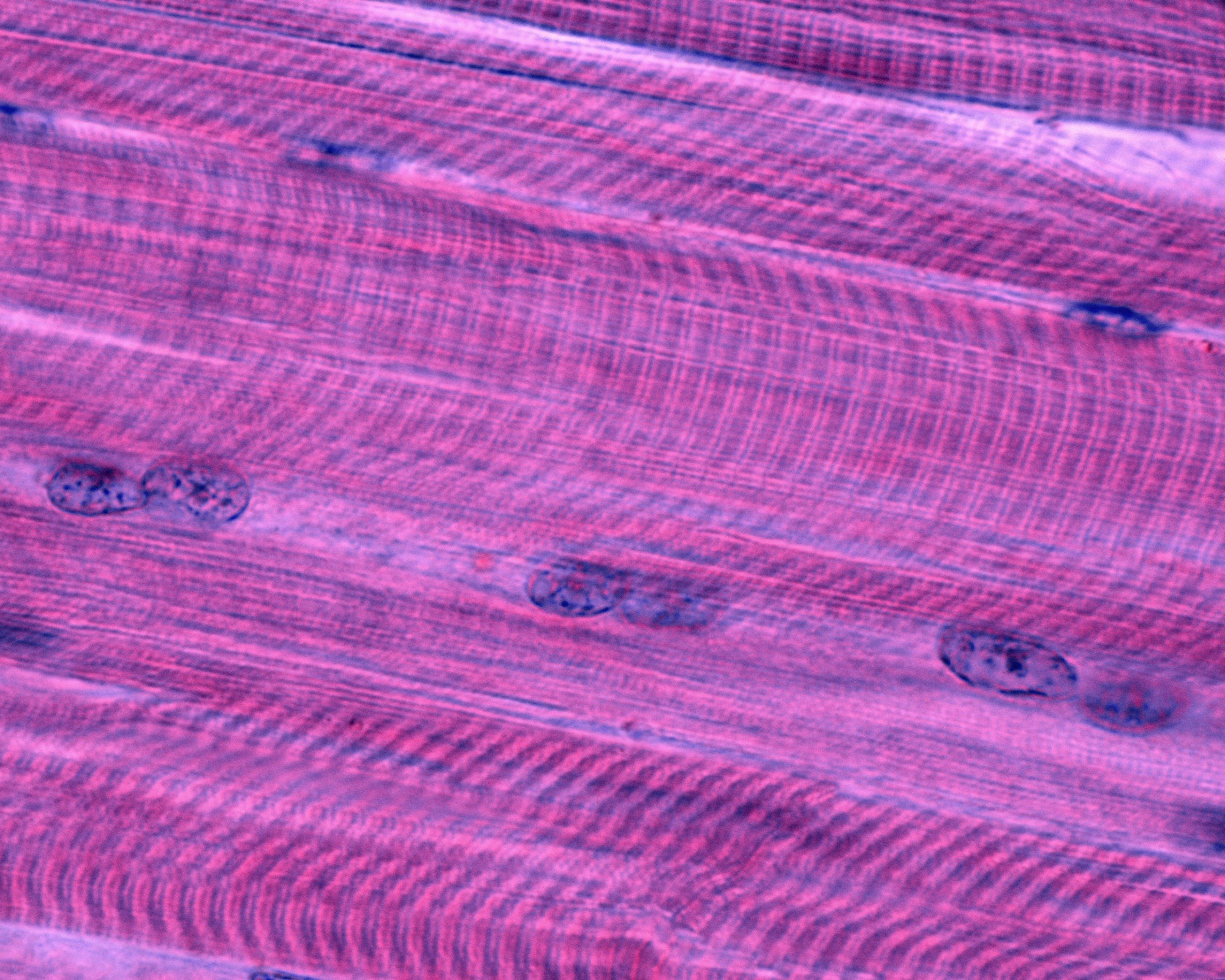
By Dr. Chinta SidharthanReviewed by Lexie CornerDec 13 2024The alignment of cells within tissues is critical for maintaining their function, especially in skeletal muscle, where proper orientation is essential for effective force generation.

Image Credit: Jose Luis Calvo/Shutterstock.com
A recent study published in Scientific Reports explored the use of ultrasonic vibrations to control the alignment of myotubes, offering a non-invasive method to manipulate cellular arrangement. By applying ultrasonication to cultured mouse myoblast cells, the researchers successfully promoted myotube alignment and differentiation, showing potential for advanced techniques in tissue engineering and regenerative medicine.
Background
The proper alignment of cells in skeletal muscle is necessary for its contractile function. However, traditional methods for culturing muscle tissues often fail to achieve the required level of alignment, posing challenges for tissue engineering. Various approaches, including chemical, electrical, and mechanical stimuli, have been tested, but each has limitations in terms of scalability and biological compatibility.
Ultrasonication, a technique that uses sound waves to manipulate cells, has emerged as a promising alternative. While ultrasound-based methods have been used to influence cell adhesion and growth patterns, their potential to specifically align myotubes for functional muscle tissue development has not been fully explored.
The current study
This study used ultrasonication to manipulate the alignment of C2C12 myoblasts and evaluate its impact on their differentiation into myotubes. C2C12 cells, a mouse myoblast cell line, were cultured in a medium containing fetal bovine serum under standard conditions and later differentiated using a reduced-serum medium.
The experimental setup included a culture dish with a glass plate bonded to a piezoelectric transducer, which generated concentric vibrations at 89 kHz. Silicone oil was applied to improve the transmission of vibrations to the culture dish.
Two ultrasonication protocols were tested. One applied continuous vibrations for 168 hours after differentiation began, while the other applied vibrations only during the first 48 hours after cell seeding, before differentiation. The vibrational amplitude was carefully controlled, ranging from 5 to 30 peak-to-peak voltage (Vpp).
The researchers assessed the effects of ultrasonication on cell adhesion, orientation, and alignment using phase-contrast microscopy. They measured cell alignment using two-dimensional Fourier transform analysis, which revealed changes in the directionality of the cells.
To explore the molecular impact of ultrasonication, real-time polymerase chain reaction (PCR) was used to measure the expression levels of muscle differentiation-related genes. Additionally, immunostaining against myosin-heavy chain provided further insights into differentiation. Statistical analysis of cell orientation and alignment confirmed that ultrasonic vibrations influenced the cellular arrangement.
Major findings
The results indicated that ultrasonication had a significant effect on the alignment and differentiation of C2C12 cells into myotubes. Myotubes cultured without ultrasonication showed random orientations, while those subjected to ultrasonic vibrations demonstrated a distinct circumferential alignment, particularly under procedure 2.
This protocol, which applied ultrasonication during the first 48 hours after cell seeding, resulted in a smaller deviation in alignment angles compared to procedure 1, where ultrasonication was applied for 168 hours after differentiation began.
Gene expression analysis showed that ultrasonication increased the expression of key differentiation-related markers, with significantly higher levels observed under higher vibrational amplitudes (20–30 Vpp) in procedure 2. These markers included myoblast determination protein 1 (Myod1), myogenin, myocyte enhancer factor 2C (Mef2c), extracellular signal-regulated kinase 5 (ERK5), Kruppel-like factor 2 (Klf2), and muscle creatine kinase (mCK).
Immunostaining revealed that a greater proportion of myotubes expressed skeletal muscle myosin heavy chain, with stronger signal intensity in the ultrasonicated samples, indicating enhanced differentiation and improved structural organization.
Quantitative analysis showed that vibrational amplitude played a critical role, with higher amplitudes leading to better alignment. Myotubes cultured with 30 Vpp ultrasonication exhibited the highest alignment and fusion rates, suggesting optimal conditions for improving cellular organization and development.
The study also showed that the alignment induced by ultrasonic vibrations corresponded to the vibrational displacement pattern on the culture dish, confirming a direct relationship between the mechanical stimuli and the cells' response.
The findings demonstrate that ultrasonication is a promising tool for promoting myotube alignment and differentiation, with potential applications in tissue engineering and regenerative medicine.
Conclusions
The study highlighted the potential of ultrasonication to align myotubes and enhance their differentiation, presenting a non-invasive and scalable approach for tissue engineering. By fine-tuning vibrational amplitude and timing, the researchers were able to achieve substantial alignment and increase the expression of muscle differentiation markers.
These results open the door to further development of ultrasound-based techniques for creating functional skeletal muscle tissues, which could help meet critical demands in regenerative medicine and bioengineering.
Journal reference
Hashiguchi, R., Ichikawa, H., Kumeta, M., Koyama, D. (2024). Control of myotube orientation using ultrasonication. Scientific Reports, 14(1), 25737. DOI:10.1038/s4159802477277x, https://www.nature.com/articles/s41598-024-77277-x