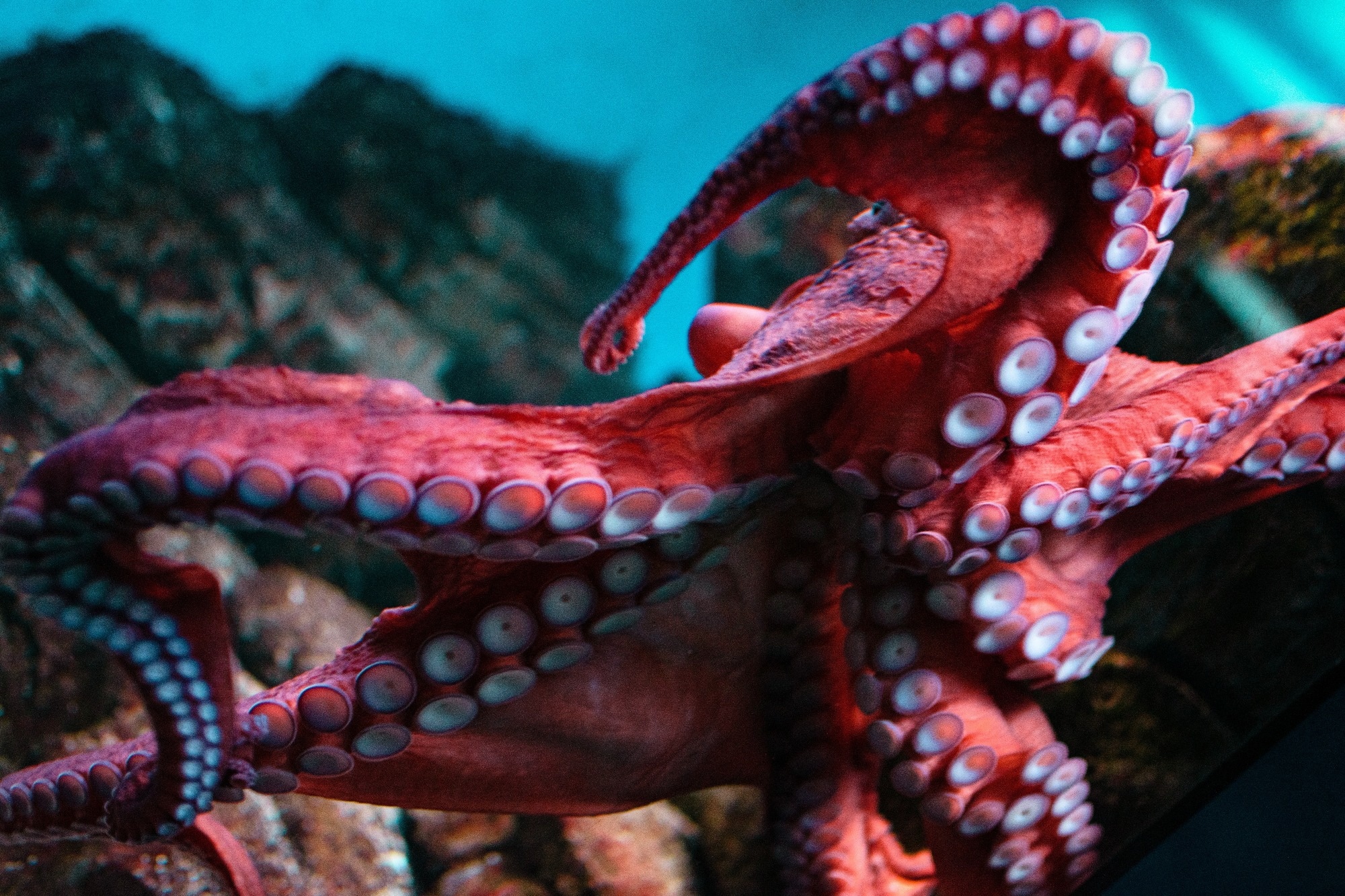
The arms of cephalopods such as octopuses are a marvel of nature. They combine flexibility and precision without a skeletal framework. These appendages enable complex movements and sensory exploration and are supported by a unique and extensive nervous system.
In a recent study published in Nature Communications, scientists from the University of Chicago investigated the axial nerve cords of cephalopods, exploring their cellular and molecular structure. The research revealed that these nerve cords are segmented and form distinct modular units along the arms, which are associated with sucker function and motor control.
 Study: Neuronal segmentation in cephalopod arms. Image Credit: Azrialette/Shutterstock.com
Cephalopod Arms
Specialized neural architectures often support movement and dexterity in soft-bodied animals. Cephalopods, which include octopuses, squids, and cuttlefish, exhibit extraordinary neural complexity, particularly in their arms. These appendages contain a high density of neurons, outnumbering even the central brain.
The axial nerve cord lies at the core of the arm's motor and sensory integration and is crucial for controlling intricate movements and sucker dynamics.
However, although previous research has described the general anatomy and neural richness of cephalopod arms, the detailed organization and functionality of their nervous system remain poorly understood.
Furthermore, segmentation, a well-known feature in some animal groups, has not been extensively studied in cephalopod neural systems.
Understanding how segmentation influences sensorimotor control in cephalopod arms may reveal fundamental principles of neural organization and help address gaps in knowledge about the evolution and function of modular nervous systems in soft-bodied animals.
About the Study
The present study examined the axial nerve cords of cephalopods to determine their structural and functional organization. The researchers used wild-caught specimens of the octopus species Octopus bimaculoides and the squid Doryteuthis pealeii, ensuring strict compliance with animal care guidelines.
Adult octopuses were housed in controlled environments, anesthetized, and dissected for tissue collection. The arms were sliced into sections for detailed analysis.
Histological techniques were employed to visualize the axial nerve cords. The researchers used stains such as acetylated alpha-tubulin to highlight neural structures and phalloidin to label actin filaments. Additionally, in situ hybridization was performed to identify molecular markers specific to sensory and motor neurons.
Imaging of stained sections was conducted using advanced microscopy, including confocal and 2-photon setups, for high-resolution visualization. The team performed neural tracing by injecting fluorescent dyes into axial nerve cord sections and tracking axonal projections to adjacent tissues such as suckers.
To study the relationship between axial nerve cord segmentation and sucker function, specific regions of the nerve cord were also examined for patterns of spatial organization.
The researchers also analyzed the arrangement of nerve exits in the septa, which are the gaps between segments in the arms of the octopus that allow nerves and blood vessels to connect to the muscles and suckers and correlated their placement with a spatial map linking nerve modules to individual suckers.
Major Findings
The results revealed that the axial nerve cords of cephalopods exhibit distinct segmentation, a feature critical for motor and sensory control. Each segment corresponds to a sucker and is organized into modular units containing sensory and motor components. This arrangement enables precise coordination of sucker movements.
The axial nerve cord segmentation was also found to be characterized by repeated cellular layers around the neuropil or network of axons, dendrites, and glial cells, with distinct exit points for the nerves targeting the suckers.
These nerves formed a spatial map, which the researchers called “suckerotopy,” that connects specific axial nerve cord segments to individual suckers.
This map was also found to facilitate intra-sucker and inter-sucker communication, enabling coordinated actions across the arm. Moreover, differences in segment density and width were observed along the arm's length, reflecting variations in neural resource allocation.
The comparative studies also revealed that segmentation is prominent in sucker-laden arms and tentacle clubs but less distinct in stalks lacking suckers. This suggested that axial nerve cord segmentation is an adaptation associated with the demands of flexible, sucker-equipped appendages.
Additionally, the study demonstrated asymmetry in axial nerve cord segment width, with more neural territory allocated to the external sucker side. Despite this, the sensory epithelium of the sucker showed even innervation, indicating a functional topographic organization.
These findings highlighted the critical role of segmentation in facilitating the cephalopod’s advanced motor and sensory abilities.
Conclusions
Overall, the findings linked the segmented organization of axial nerve cords in cephalopod arms to the precise control of sucker movements.
By revealing the spatial mapping and modular structure of the axial nerve cords, the study also provided insights into the neural basis of motor control in soft-bodied animals.
These findings not only further our understanding of cephalopod biology but also contribute to broader discussions on neural segmentation and its evolutionary significance in flexible appendages.