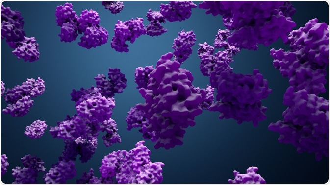
Protein localization at the right time at the right place is very important to gain access to appropriate molecular interaction partners. The proper subcellular protein localization is pivotal for providing the physiological context for protein function. Proteins perform their biological functions within the spatiotemporal context of an intact cell.

Image Credit: Design_Cells/Shutterstock.com
Regulation of protein localization
Rapid changes in local protein function in cells could be achieved via specifically redirecting the localization of the pool of existing protein. Eukaryotic cells have developed sophisticated targeting pathways to direct proteins to the appropriate cellular locations.
There are two main pathways of protein localizations: nuclear and mitochondrial pathways. The nucleus contains the majority of the cell's genetic material and helps in keeping the genetic information physically separated from other cellular functions such as transcription and translation processes. This ensures that RNA processing (including splicing, capping, cleavage/polyadenylation, and folding) will be run with no risk of intermediates in the process being translated and interfering with the normal functions of the cell.
The nuclear envelope, which is a double-bilayer membrane contiguous with the endoplasmic reticulum, separates the nucleoplasm from the cytoplasm. Large multiprotein complexes called nuclear pore complexes control the transport through the envelope. The nuclear pore complexes, comprised of a class of proteins termed nucleoporins, can form a large macromolecular conduit between the cytoplasm and the nucleoplasm.
Directionality is conferred on the transport of macromolecular into and out of the nucleus via control of binding and release of cargo, which is dependent on the small GTP-binding protein/GTPase, Ran.
Nuclear genes produce the majority of mitochondrial proteins. Then, they will be imported into mitochondria. In fact, mitochondrial protein import is a highly complex process due to the many functional locations in the mitochondrion, and its bacterial origin. Protein import starts with the mitochondrial protein binding to and moving through the translocase of the outer membrane complex in either an unfolded conformation or α-helical.
Mechanisms governing protein localization
The protein distribution among cellular compartments depends on four main factors: (1) the number and types of localization protein signals, (2) the concentration of freely diffusing molecules, (3) the relative strength of each signal, and (4) the activity and concentration of localization signal receptors. The localization of a specific protein is affected by the first three factors, while the fourth factor permits global control of total classes of proteins.
Modifying the binding affinity of the localization signal for protein corresponding receptor is one of the most common methods by which cell controls the distribution of protein. Post-translational modification of the cargo protein in or near the localization signal achieves this affinity modulation. This type of mechanism usually involves serine/threonine phosphorylation, however may also occur through lysine sumoylation, tyrosine phosphorylation, or lysine acetylation. These modifications interfere with localization receptor binding.
Signal addition is another common method of adjusting protein localization. By necessity, signal addition mandates pores to handle multiple folded proteins, like the peroxisomal pore, ciliary boundary, and the nuclear pore. Signal addition is not applicable for a system that typically needs an unfolded protein, like the endoplasmic reticulum or mitochondrial translocons.
Signal addition facilitates the protein localization to be indirectly regulated by the localization and levels of its binding partner, which has a comparable role to import receptors. Signal masking is a very efficient method to prevent protein transport by burying the signal. Hiding the signals may either be by another protein or ligand, or signals may be obscured by an intramolecular interaction.
Protein sequestration within a compartment happens if the number of binding sites within the compartment remarkably decreases the protein mobility so the export rate from the compartment is decreased. Tethering, or anchoring, is a unique form of sequestration where the protein is embedded in or bound to an essentially immobile component with respect to other components, like the chromatin, cytoskeleton, or plasma membrane. Tethering decreases the soluble pool available to be acted on by transport receptors.
Cells have evolved countless approaches for the regulation of protein localization and consequently function. Most of these mechanisms permit quick reversible regulations like post-translational modification and conformational changes, while few need novel synthesis for changing of localization.
High Resolution Mapping of Protein Localization in Mammalian Brain
Sources:
- Hung, Mien-Chie, and Wolfgang Link. "Protein localization in disease and therapy." Journal of cell science 124.20 (2011): 3381-3392. https://doi.org/10.1242/jcs.089110
- Nakai, Kenta, and Minoru Kanehisa. "A knowledge base for predicting protein localization sites in eukaryotic cells." Genomics 14.4 (1992): 897-911. https://doi.org/10.1016/S0888-7543(05)80111-9
- Bauer, Nicholas C., Paul W. Doetsch, and Anita H. Corbett. "Mechanisms regulating protein localization." Traffic 16.10 (2015): 1039-1061. https://doi.org/10.1111/tra.12310
- Luk, Edward, et al. "Manganese activation of superoxide dismutase 2 in the mitochondria of Saccharomyces cerevisiae." Journal of Biological Chemistry 280.24 (2005): 22715-22720. https://doi.org/10.1074/jbc.M504257200
- Yogev, Ohad, Sharon Karniely, and Ophry Pines. "Translation-coupled translocation of yeast fumarase into mitochondria in vivo." Journal of Biological Chemistry 282.40 (2007): 29222-29229. https://doi.org/10.1074/jbc.M704201200
- Hill, Kerstin, et al. "Tom40 forms the hydrophilic channel of the mitochondrial import pore for preproteins." Nature 395.6701 (1998): 516-521. https://doi.org/10.1038/26780
- Künkele, Klaus-Peter, et al. "The preprotein translocation channel of the outer membrane of mitochondria." Cell 93.6 (1998): 1009-1019. https://doi.org/10.1016/S0092-8674(00)81206-4
- Hoelz, André, Erik W. Debler, and Günter Blobel. "The structure of the nuclear pore complex." Annual review of biochemistry 80 (2011): 613-643. https://doi.org/10.1146/annurev-biochem-060109-151030
Further Reading
Last Updated: Nov 23, 2021