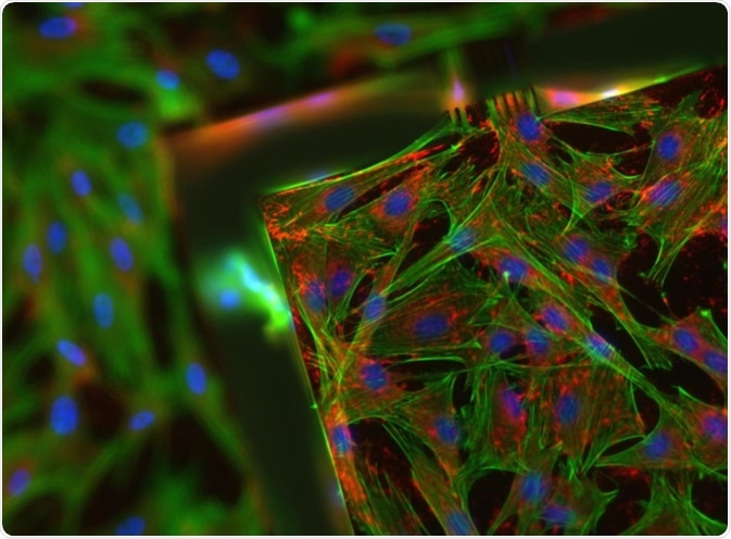
Fluorescence microscopy is a common laboratory tool used to investigate the physiology of different cells. Specific wavelengths of light are shone onto cells in a sample, where the light is absorbed by molecules called fluorophores.
 Image Credit: Virginie Thomas/Shutterstock.com
There are many types of fluorophores, all of which absorb light and re-emit light of a longer wavelength, visible as a different color. Other light present is filtered out by the fluorescence microscope, and the re-emitted light is shown to the viewer.
It is important to prevent cell death during fluorescence microscopy, as samples may need to be studied for relatively long lengths of time and dying cells may skew experimental results.
Phototoxicity and photobleaching
When the fluorophores in the sample are excited by the light source, negative effects can occur. These are called phototoxic effects.
Naturally occurring fluorophores like flavins and porphyrins are degraded when they absorb light, producing reactive oxygen species (ROS) such as radicals and peroxides. This process is called photobleaching.
ROS are the main negative factor in phototoxicity. ROS can damage DNA and RNA, and oxidize lipids and proteins, affecting the essential metabolism of the cell.
Some fluorophores are more phototoxic than others. For example, protein fluorophores are less toxic as they contain a structure called a β-barrel, which keeps any negative factors produced by photobleaching contained within itself. The damage to a cell in fluorescence microscopy, therefore, depends on the type of cell, as well as the microscope instrumentation.
Choose fluorophores that produce excitation in the far red range, avoid those parts of the spectrum that cause autofluorescence, and increase the distance on the spectrum between different fluorophores to limit bleed-through.
How to limit photobleaching
There are many different adjustments that can be made to fluorescence microscopy to reduce the amount of fluorophore breakdown. The main aim is to limit the light exposure to the fluorophores, and the intensity of the light source, without reducing the quality of the output too much.
The precise wavelengths of the light source and the emission filters involved should be suitably designed for the specific fluorophores in use, in order to reduce any unnecessary excess light from producing photobleaching. Reducing any background light in the room also reduces this.
A more modern way to reduce photobleaching in cells is using pulsed illumination. This is where the subject cells are lit up in pulses with extremely short intervals of darkness, rather than constant illumination. This gives the fluorophores time to return to a relaxed state and stops the production of ROS.
Shutter speed also affects photobleaching
The shutter speed limits the amount of light that reaches the sample. Therefore increasing this shutter speed will decrease photobleaching and lead to a longer lifespan of the sample. New microscopy technologies tend to implement faster-motorized shutters which can open and close milliseconds before and after an image is taken.
A different approach to this is using an LED light source, which gets rid of the need for a shutter at all because LEDs can be switched on and off instantly.
Controlling the cellular microenvironment
One of the major conditions that must be maintained is the temperature. The organism from which the sample arises must be maintained at its ideal temperature in order to keep cell viability. This can be maintained using stage mounted incubation chambers, or using heated air that is blown across the sample.
A technique called oxygen scavenging can also be used. The most common oxygen scavenging system uses two enzymes; glucose oxidase and catalase. These enzymes oxidize glucose within the cell, which decreases the amount of oxygen present and reduces the production of ROS.
When using this oxygen scavenging system, it unfortunately also produces gluconic acid. This can interfere with proteins in the cell and is potentially harmful to the sample.
Further Reading
Last Updated: Feb 1, 2021