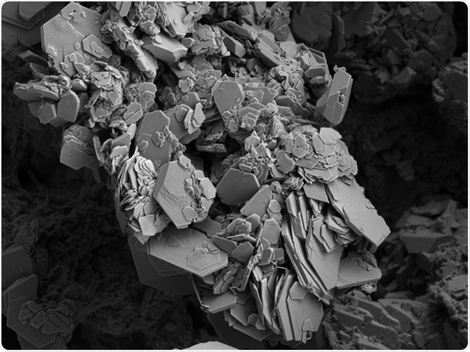
Electron microscopy has been used in various scientific disciplines from biomedical research involving tissues and cells, forensic research for trace evidence, as well as the analysis of nanomaterials in food. Electrons that act as the source of the radiation, have very short wavelengths that generate electron microscopy images with high resolution.

Electron Microscopy Image. Image Credit: Ravenash/Shutterstock.com
Principles of Electron Microscopy Technique
Unlike light microscopes that rely on a light beam focusing on an object to examine the details of a material, electron microscopes use the interaction of an electron beam with the material to examine the morphology and composition in high detail.
The image of the object can be magnified by passing the beam of electrons through the object and a series of lenses, and the scattering of the electrons in the object then results in the image being viewed.
Electromagnetic and electrostatic lenses consisting of a solenoid, a wire coil wrapped on the outside of the microscope tube, is used in all different types of electron microscopes. Manipulation of the current through the objective lens, the image of the specimen can be focused and the current value through the projector and intermediate lens gives its magnification.
The two most frequent techniques of electron microscopy are scanning electron microscopy (SEM) and transmission electron microscopy (TEM). SEM relies on emitting electron beam on the surface of the sample and detecting the secondary electron signals, thus producing images of high resolution. While TEM allows the electron beam to penetrate the sample, giving a projection view of thin samples.
Components of An Electron Microscope
The common parts that can be found within an electron microscope consist of an electron gun, electromagnetic lens, specimen holder, an image viewing system as well as a recording system. A heated tungsten filament acts as the electron gun generating electron beams that interact with the sample.
The electromagnetic lenses consist of three lenses with different functions: the condenser lenses, the objective lens, and the projector lenses. The electron beam can be focused on the specimen by a condenser lens, and the electrons are formed into a thin beam by the second condenser lens. The objective lens then allows the passing down of an electron beam from the sample, forming an intermediate image that is magnified. The final image is then generated by the projector lenses.
The specimen that is examined is held by the specimen holder that is made up of a collodion supported by a metal grid or a very thin carbon film. A fluorescent screen is used to visualize the projected final image and this image can then be recording using a camera located beneath the screen.
Sample Preparation
The sample preparation for SEM depends on which sample will be examined. Samples containing metals generally do not require any sample preparation as metals conduct electricity when electrons are bombarded, and nonmetal samples are prepared using a sputter coating system.
The nonmetal specimen is coated with gold as a common conducting material layer from an electric field and argon gas, and positively charged ions are generated from the removal of an electron from argon by the electric field. Water residuals must also be removed when utilizing the conventional SEM technique due to its vaporization when in a vacuum and can obliterate the quality of the image generated.
On the other hand, the TEM sample preparation process involves the steps including processing, rinsing, post-fixation, dehydration, and infiltration. As TEM is commonly used to analyze biological matrices, glutaraldehyde is used to fixate the protein molecules to their neighboring molecules by covalent cross-linking to preserve and stabilize the sample from deteriorating. The samples are then washed with a buffer to maintain the pH and prevent tissue fixation as a result of too much acidity.
Post fixation is then done to provide stability, preventing coagulation by alcohol during dehydration, and giving a contrast of the different structures during imaging. An organic solvent is then used in the dehydration process to replace the water in the tissue sample as epoxy resin that will be used in the next steps are immiscible with water.
The cells are then penetrated by epoxy resin during the infiltration step and flat molds are used for embedding. The sample is then placed in the oven at 60 degrees overnight to let the resin set. The samples are then cut into thin sections of 30 to 60 nm, collected to a copper grid, and examined under the TEM.
2 The Principle of the Electron Microscope
Limitations of Electron Microscopy
Although given the ability for high magnification and high resolution, there are some limitations with electron microscopy. Electron microscopes are usually expensive to build and require proper maintenance for their functionality. It is also highly sensitive to vibration, requires training for the user to operate the microscope, and live specimens cannot be examined by an electron microscope.
Applications of Electron Microscopy
Various medical science laboratories have used electron microscopy to study bacteria, viruses, and other microorganisms to aid research in different diseases. SEMs are also often utilized for test and failure analysis for material science research, examining superconductors in high temperatures, alloy strength, and nanotubes, ultimately impacting the electronics to the aerospace industry.
SEMs have been used to aid forensic investigations by the analysis of paint particles from car chips, banknote authenticity, gunshot residues, and bullet markings. By the magnification that SEMs provide, the origin of these evidence types can be determined, and conclusions of the investigation can be drawn for legal purposes.
References:
- University of Massachusetts Medical School. 2020. What Is Electron Microscopy? - UMASS Medical School. [online] Available at: <https://www.umassmed.edu/cemf/whatisem/#:~:text=Electron%20microscopy%20(EM)%20is%20a,cells%2C%20organelles%20and%20macromolecular%20complexes.> [Accessed 18 December 2020].
- Picó, Y., 2018. Safety Assessment and Migration Tests. In Nanomaterials for Food Packaging (pp. 249-275). Elsevier.
- Titus, D., Samuel, E.J.J., and Roopan, S.M., 2019. Nanoparticle characterization techniques. In Green Synthesis, Characterization and Applications of Nanoparticles (pp. 303-319). Elsevier.
- Vlab.amrita.edu. 2020. Transmission Electron Microscopy (Theory): Cell Biology Virtual Lab I: Biotechnology And Biomedical Engineering: Amrita Vishwa Vidyapeetham Virtual Lab. [online] Available at <http://vlab.amrita.edu/?sub=3&brch=187&sim=784&cnt=1> [Accessed 18 December 2020].
- ATA Scientific. 2019. The Application and Practical Uses of Scanning Electron Microscopes. [online]. Available at: https://www.atascientific.com.au/sem-imaging-applications-practical-uses-scanning-electron-microscopes/ [Accessed 18 December 2020].
Further Reading