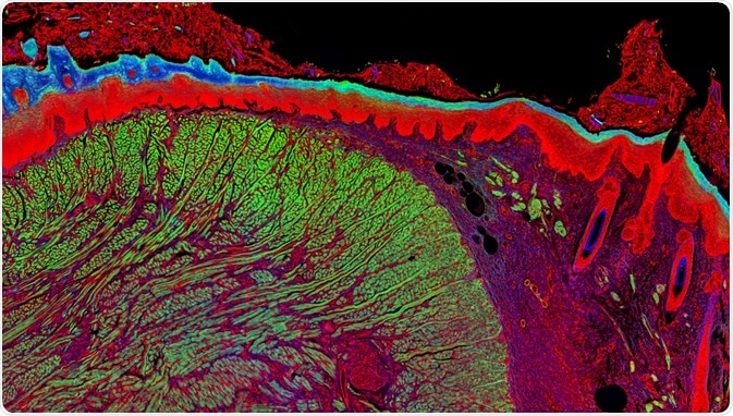
Fluorescence microscopy is a major tool used by scientists across many disciplines ranging from cell biology and histopathology to material sciences.

Fluorescence Microscopy. Image Credit: Micha Weber/Shutterstock.com
Stoke’s shift
Fluorescence microscopy is dependent upon the amount of light energy, otherwise known as photons, that are absorbed by a specified fluorescent indicator. Typically, the fluorescent indicator will emit a lower amount of light and therefore have a longer wavelength as compared to that of the excitation light, which is the light energy that is being absorbed. Blue light, which has a shorter wavelength, therefore has higher energy as compared to red light, which has a longer wavelength.
This phenomenon, otherwise known as the Stokes shift or Stoke’s Law, occurs due to the energy loss that occurs during the absorption of light energy by the indicator. As the value of Stoke’s shift increases, the ease of separating excitation light from emission light through combinations of the fluorescence filter also increases. Since the emission of a fluorophore is usually lower than both the wavelength and magnitude of the excitation light, the emission spectral curve is a mirror image of the excitation curve.
Alexa Fluor 555, for example, absorbs light in the yellow-green region while producing light that is in the yellow-orange emission region. Maximum fluorescence intensity is therefore achieved through the use of a fluorophore or dye that is excited at wavelengths that are at or near the peak of the excitation curve.
Two-photon fluorescence microscopy
Fluorescent indicators are capable of absorbing single or multiple photons while simultaneously only emitting one photon. Otherwise known as two-photon or multiphoton fluorescence microscopy, this process requires the use of a highly specialized and high-powered pulsed laser to ensure that two absorbed photons arrive simultaneously. Through this form of fluorescence microscopy, the two photons that are absorbed at the same time have an energy that is less than that of the emitted photon.
As a result, red light, which is a longer-wavelength type of light, can be used to generate green light. The advantage of using red light over blue light, which is a shorter-wavelength light, is a deeper penetrance of the light into tissues, a lower amount of scattering of the light as well as a reduced likelihood that the light will damage the cells being analyzed.
The design of a fluorescence microscope
A typical epi-fluorescence microscope will be capable of performing both transmitted and reflected fluorescence microscopy. Excitation light, which is emitted from the arc-discharge lamp or another light source, is directed through a wavelength selective excitation filter and ultimately reflected on the surface of a dichromatic mirror or beam splitter.
The excitation light will be of a specific wavelength, most often of which includes those within the ultraviolet, blue, or green regions of the visible spectrum. Once reflected by the mirror, the excitation light will pass through the microscope objective to directly bathe the sample with light.
If the sample fluoresces, the emission light is gathered by the microscope objective and travels back through the dichromatic mirror to be filtered by the emission filter. The emission filter, which is also known as the barrier or suppression filter, blocks any unwanted excitation wavelengths, ultimately allowing for the desired and longer wavelengths to be passed onto the eyepiece or detector.
Conditions that affect fluorescence emission
Typically, the initial radiation of fluorescence emission on a sample will not affect the emission intensity. However, problems begin to arise upon subsequent re-radiation of the samples. Fading, for example, is a term used to describe a reduction of fluorescence emission intensity.
Photobleaching
Photobleaching, which is a specific type of fluorescence fading, is the irreversible decomposition of the fluorescent molecules in their excited state as a result of their unwanted interaction with oxygen before their emission.
Although photobleaching is typically an unwanted occurrence in fluorescence microscopy imaging, it can be exploited for some specific biological studies. Fluorescence recovery after photobleaching (FRAP), for example, utilizes photobleaching to investigate the diffusion and motion of macromolecules, whereas fluorescence loss in photobleaching (FLIP) monitors the reduction in fluorescence emission to determine the mobility and other dynamics of biological cells.
Quenching
Another type of fading that can occur in fluorescence imaging is quenching, which is a process in which the fluorescence intensity gradually decreases. A variety of different mechanisms are responsible for quenching, such as non-radiative energy loss as well as exposure to certain substances including oxidizing agents, salts, heavy metals, or halogen compounds.
Types of fluorescence microscopes
A basic wide-field fluorescence microscope is often used by the modern cell biologist. Typically, this instrument will provide excitation light through a mercury or xenon high-pressure bulb; however, more advanced versions of this type of fluorescence microscope will use bright single-wavelength light-emitting diodes (LEDs). As compared to the traditional mercury or xenon bulb, LEDs offer longer life, eliminate the need for shutters by providing fast switching and also ensure optimal wavelength control.
Another popular fluorescence microscope is the laser-scanning confocal microscope (LSCM). As compared to the wide-field fluorescence microscope, the LSCM has a bright point source, such as a laser, for its excitation source, as well as a sequential scanning method as the illumination source. An additional difference between the LSCM and the wide-field microscope is that a photomultiplier tube (PMT) is used to detect emitted light intensity.
The confocal microscope is a highly useful imaging tool, as it is capable of rejecting any light that is out-of-focus from altering the integrity of the image. While this advantage can be attributed to some of the aforementioned differences that exist between confocal microscopes and wide-field fluorescence microscopes, it is largely due to the pinhole aperture within this imaging device.
More specifically, the pinhole aperture ensures that the only light that will reach the detector will come from the confocal point from which the excitation light was focused on in the sample being analyzed. As the laser and pinhole remain completely still throughout the imaging process, their area of focus is optically moved across the specimen by a pair of oscillating mirrors.
Further Reading