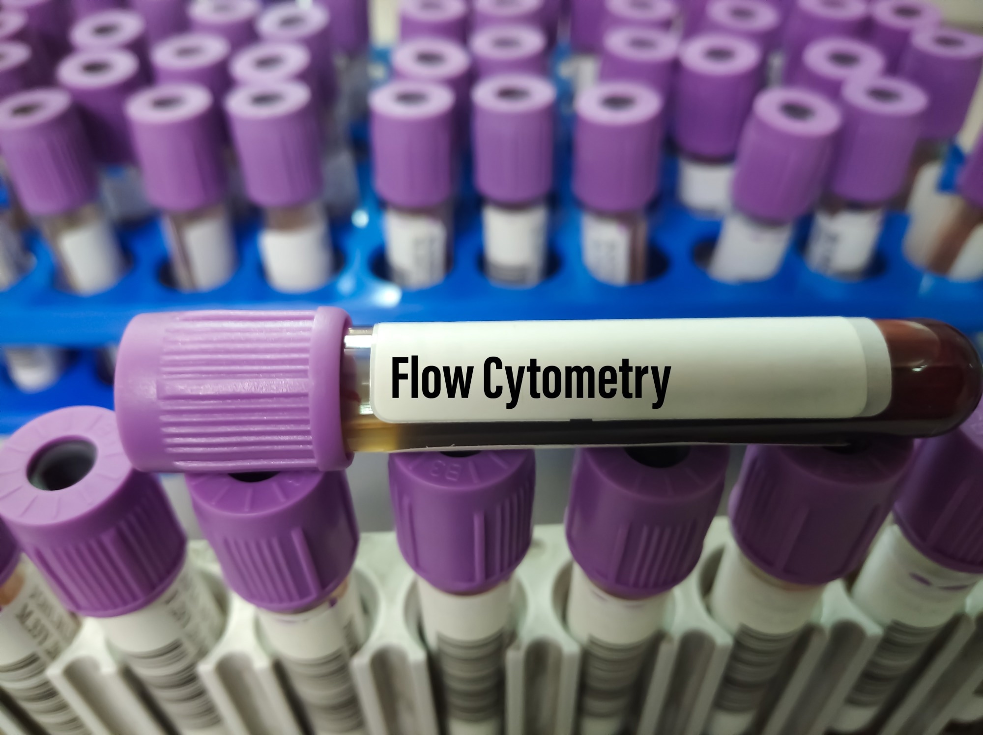
Flow cytometry detects and measures the chemical, fluorescent, and physical characteristics of cells and particles passing in a fluid stream. This technique analyzes cells by passing them through laser beams and then collecting the scattered/emitted light.1
 Image Credit: Babul Hosen/Shutterstock.com
Flow cytometry plays a critical role in immunology for immune profiling, oncology for cancer cell marker identification, drug discovery for cell-based assay screening, and clinical diagnostics for detecting diseases like leukemia and infections.1
Image-based flow cytometry (IFC) is an advanced method for thoroughly quantifying and analyzing cells in intricate samples, enabling a holistic understanding of biological processes. The method combines computational analysis, optical microscopy, and flow cytometry. IFC facilitates investigations into various processes, such as disease-related alterations, cell differentiation, and cell-cell interactions.2
How is Flow Cytometry Used in Biotechnology?
The Role of AI in Image Analysis for Flow Cytometry
Artificial intelligence (AI) and machine learning (ML) are transforming flow cytometry, specifically IFC, by significantly improving data analysis. AI’s learning capacity and adaptability could play a major role in knowledge mining from the IFC-generated high-dimensional data, resulting in more accurate analysis.3-5
The application of ML algorithms has been extensive, specifically in rapid image processing as a segmentation tool in advanced IFC systems. Deep learning enables semantic segmentation of complex structures/cells by assigning a label/category to every pixel within the sample image.3-5
Similarly, convolutional neural networks (CNN) are less dependent on manually chosen segmentation parameters and could generate higher-quality results. Studies reported that general-purpose ML methods could address image artefacts and aberrations caused by acquisition system imperfections.3-5
Studies have used deep learning and IFC to distinguish clinically relevant red blood cell morphologies linked with cell storage lesions. In commercial IFC, ML algorithms have been used for label-free leukemia monitoring, white blood cell identification, and label-free cell-cycle identification.3-5
Although conventional feature extraction and classifiers like gradient boosting (GB), random forest (RF), support vector machine (SVM), k-nearest neighbors (KNN), and AdaBoost were used in the early stages, deep learning-based algorithms like PyramidNet, NasNet, VGG, AlexNet, and conventional CNN have later gained attention as they could be optimized to realize high prediction accuracies.3-5
A work recently published in Computacion y Sistemas introduced deep learning-based classification and segmentation of sperm head and flagellum for IFC. The quantitative results from the ResNet50 classification displayed exceptional performance, realizing a 0.99 F1 score/near-perfect classification accuracy. Additionally, an average dice coefficient of 0.81 was achieved using the U-Net segmentation.2
How AI Enhances Flow Cytometry
AI enhances flow cytometry by improving accuracy and data processing, enabling real-time insights, and uncovering hidden cellular patterns. It reduces human error and accelerates the analysis of large, complex datasets.3-5
Improved Accuracy and Precision
AI improves precision in flow cytometry, resulting in more accurate and reliable results compared to traditional methods. Studies have shown that two-stage models like faster-region CNN and one-stage models like single-shot detectors provide better performance on detection accuracy and speed while detecting cells infected by malaria parasites and neural cells, respectively.3-5
Other algorithms used that display over 90% accuracy include GB, RF, AlexNet, GoogleNet, DenseNet, VGG, MobileNetV2, CNN, NasNet, ResNet, linear SVM, KNN, AdaBoost, and SVM. These algorithms are primarily used for cell imaging analysis, leukemia monitoring, and white blood cell identification.3-5
Enhanced Data Processing
AI enables faster analysis of high-dimensional, large-scale datasets. Specifically, deep learning excels in analyzing IFC data, offering both high sensitivity and the speed required to handle its high-throughput nature.3-5
The successful application of this algorithm has significantly advanced high-throughput cell analysis. For instance, 3D IFC has been successfully performed to reveal liver endothelial cell and hepatic stellate cell morphology at a single-cell resolution.
A combination of transmission and side-scattered single-cell images of liver cells with AI was used to provide a staging system of nonalcoholic steatohepatitis (NASH) progression.3-5
Real-time Insights
AI provides real-time insights, allowing for immediate classification and detection of cell populations during analysis. This capability accelerates decision-making and enhances the ability to monitor cellular dynamics in real-time.3-5
For instance, deep cytometry, a recent deep learning implementation, enables non-supervised, real-time cell sorting and eliminates the need for image reconstruction.
Hence, AI ensures timely, accurate results, crucial for applications such as disease monitoring and therapeutic development.3-5
Novel Discoveries
AI could reveal previously hidden patterns in cellular morphology and behavior. This ability to uncover novel insights leads to a deeper understanding of cellular dynamics and can drive breakthroughs in areas like disease research and personalized medicine.3-5
A.I. in flow cytometry analysis - C2S Innovation Insights video series
Applications of AI-Driven Flow Cytometry
AI-driven flow cytometry has revolutionized various fields, offering enhanced precision and speed in analyzing complex cellular data.6-9
Cancer Research
AI-driven flow cytometry enhances cancer research by improving the detection of rare cancer cells. It also aids in characterizing tumor heterogeneity, offering deeper insights into cancer progression and treatment response.6
For instance, AI and flow cytometry-based approaches to diagnostic markers have been developed for B-cell non-Hodgkin lymphomas (B-NHL). Mature B-cell lymphoid neoplasms consist of a diverse and heterogeneous group of malignancies characterized by variations in morphology, phenotype, genotype, and aggressiveness. ML algorithms could be applied to a large dataset of B-NHL immunophenotypes to generate a clinically applicable and robust prediction system.6
Immunology
AI-driven flow cytometry enables more precise identification of immune cell populations. This advancement supports the development of vaccines and immune therapies by providing deeper insights into immune responses and cell behavior.7
A work recently published in Diagnostics developed and validated AI-assisted flow cytometry workflow for primary immunodeficiency diseases and related immunological disorders using 379 clinical cases from 2021 and a 3-tube, 10-color flow panel with 21 antibodies.7
The AI software was fully automated, which reduced analysis time to less than 5 min per case. Using an exclusive multidimensional density–phenotype coupling algorithm, the AI model accurately classified and enumerated NK, B, and T cells, along with crucial immune cell subsets like CD8+ cytotoxic T cells, CD4+ helper T cells, and class-switched or non-switched B cells.7
Clinical Diagnostics
AI-driven flow cytometry improves clinical diagnostics by enhancing disease detection and monitoring accuracy. This technology aids in the more precise identification of conditions like leukemia and HIV, facilitating earlier intervention and better patient outcomes.8
A work recently published in Haematologica reported a novel model integrating AI with multiparametric flow cytometry to enhance the classification and diagnosis of myelodysplastic syndromes (MDS).8
An ML model was developed through an elastic net algorithm directed on a cohort of 191 patients only based on Boruta algorithm-selected flow cytometry parameters to build a reliable and simple prediction score with five parameters. The model diagnosed both low- and high-risk MDS with 92.5% specificity and 91.8% sensitivity.8
Drug Discovery
AI-driven flow cytometry accelerates drug discovery by enabling high-throughput analysis of therapeutic effects on cell populations. This technology allows for efficient screening of potential drug candidates, providing valuable insights into their impact on cellular behavior and viability.9
Evolving multi-omic and AI/ML-driven data analysis will facilitate multi-parametric, automated evaluation of cell populations, potentially minimizing the variability introduced by manual definition of cell populations and gating.9
Acoustic-Assisted Hydrodynamic Focusing in Flow Cytometry
Challenges in Implementing AI-Powered Image Analysis
Implementing AI-powered image analysis in healthcare comes with multiple challenges. Data standardization is a key issue, as inconsistent formats and quality could affect AI model performance. Integrating AI tools into existing clinical workflows requires careful planning and adaptation to ensure seamless operation.3,5
Additionally, the computational resources needed for processing large datasets can be costly and complex. Data privacy concerns also arise, especially when dealing with sensitive patient information.3,5
Moreover, the need for large, labeled datasets to train models presents logistical challenges. Validation and adoption in clinical settings are critical to ensure that healthcare professionals trust AI tools.3,5
The Future of AI in Flow Cytometry
The future of AI in flow cytometry is poised for transformative advancements, particularly through the integration of AI with multi-omics analysis and single-cell sequencing, enabling more comprehensive insights into cellular function. AI has the potential to drive fully automated flow cytometry systems, enhancing both research capabilities and diagnostic accuracy.4,5,9
This automation can streamline data analysis, reducing human error and improving efficiency. Additionally, AI will play a crucial role in expanding personalized medicine by refining precision diagnostics and facilitating targeted therapies based on individual cellular profiles.4,5,9
Conclusion
In conclusion, AI-powered image analysis is revolutionizing flow cytometry by significantly increasing its efficiency, accuracy, and application scope. By integrating AI, flow cytometry can process complex datasets faster, enhance the precision of cell detection, and uncover previously hidden patterns in cellular behavior.
In clinical settings, AI-driven flow cytometry improves diagnostic accuracy, leading to better patient outcomes. By driving innovation and expanding the potential of life sciences, AI is poised to shape the future of personalized medicine and therapeutic development.
References
- Papa, S., Ortolani, C., Fernández, P. (2023). Flow Cytometry and Its Applications to Molecular Biology and Diagnosis 2.0. International Journal of Molecular Sciences, 24(22), 16215. DOI: 10.3390/ijms242216215, https://www.mdpi.com/1422-0067/24/22/16215
- Hernández-Herrera, P., Abonza, V., Sanchez-Contreras, J., Darszon, A., Guerrero, A. (2023). Deep Learning-Based Classification and Segmentation of Sperm Head and Flagellum for Image-Based Flow Cytometry. Computación y Sistemas, 27(4), 1133-1145. DOI: 10.13053/cys-27-4-4772, https://www.scielo.org.mx/scielo.php?pid=S1405-55462023000401133&script=sci_arttext&tlng=en
- Pozzi, P., Candeo, A., Paiè, P., Bragheri, F., Bassi, A. (2023). Artificial intelligence in imaging flow cytometry. Frontiers in Bioinformatics, 3, 1229052. DOI: 10.3389/fbinf.2023.1229052, https://www.frontiersin.org/journals/bioinformatics/articles/10.3389/fbinf.2023.1229052/full
- Dimitriadis, S., Dova, L., Kotsianidis, I., Hatzimichael, E., Kapsali, E., & Markopoulos, G. S. (2024). Imaging Flow Cytometry: Development, Present Applications, and Future Challenges. Methods and Protocols, 7(2), 28. DOI: 10.3390/mps7020028, https://www.mdpi.com/2409-9279/7/2/28
- Luo, S. et al. (2021). Machine-Learning-Assisted Intelligent Imaging Flow Cytometry: A Review. Advanced Intelligent Systems, 3(11), 2100073. DOI: 10.1002/aisy.202100073, https://onlinelibrary.wiley.com/doi/full/10.1002/aisy.202100073
- Casanova, E. (2023). Flow Cytometric And Artificial Intelligence Approach To Diagnostic Markers For B-Cell Lymphomas. https://ora.uniurb.it/handle/11576/2725878
- Lu, Z. et al. (2024). Validation of Artificial Intelligence (AI)-Assisted Flow Cytometry Analysis for Immunological Disorders. Diagnostics, 14(4), 420. DOI: 10.3390/diagnostics14040420, https://www.mdpi.com/2075-4418/14/4/420
- Clichet, V. et al. (2023). Artificial intelligence to empower diagnosis of myelodysplastic syndromes by multiparametric flow cytometry. Haematologica, 108(9), 2435. DOI: 10.3324/haematol.2022.282370, https://pmc.ncbi.nlm.nih.gov/articles/PMC10483367/
- Ullas, S., & Sinclair, C. (2023). Applications of Flow Cytometry in Drug Discovery and Translational Research. International Journal of Molecular Sciences, 25(7), 3851. DOI: 10.3390/ijms25073851, https://www.mdpi.com/1422-0067/25/7/3851
Further Reading
Last Updated: Jan 2, 2025