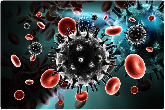
The immunopathogenic mechanisms in human immunodeficiency virus (HIV) include a sophisticated array of multifactorial and multiphasic immunopathogenic mechanisms. Although CD4+ T cells are depleted by HIV, other immune-competent cells can also be dysregulated.

Image Credit: RAJ CREATIONZS/Shutterstock.com
Dysfunction of CD4+ T Lymphocytes
The impairment of a variety of CD4+ T-cell functions is one of the early hallmarks of HIV infection. This usually occurs prior to the profound depletion in CD4+ T cells. This impairment of CD4+ T-cell functions includes autologous mixed lymphocyte reactions, T-cell colony formation, expression of interleukin-2 (IL-2) receptors, and IL-2 production.
During all stages of HIV disease, there is a decrease in T-cell proliferative responses to a range of stimuli. The response to recall antigens like influenza, Candida albicans tetanus toxoid, and Cryptococcus neoformans is the earliest T-cell proliferative response to be lost. The loss of T-cell proliferative responses to alloantigens and mitogens is usually preceded by dysfunction in responses to soluble antigens. Cooper and colleagues reported that there was a decrease in CD4+ T-cell responses to antigens and mitogens during acute HIV infection.
There is a growing conflict whether a defect in T-cell proliferative responses has a relationship with abnormalities in antigen-presenting cells. Although some researchers suggested that the defect in T-cell proliferative responses is independent of abnormalities in antigen-presenting cells, others have found that abnormalities in antigen-presenting cell function led to a secondary defect in T-cell proliferation. Meyaard and colleagues suggested that various T-cell abnormalities could be explained by the effects of HIV on antigen-presenting cells
Human Helper CD4+ T cells can be divided into two groups based on their function and profiles of secreted cytokines. The first group is T-helper-1 (TH1) cells which provide help to cytotoxic T lymphocytes (CTLs) and secrete IL-2 and interferon-γ. The second group is T-helper-2 (TH2) cells which support B lymphocytes and secrete IL-4 and IL-10.
Clerici and Shearer reported that a switch from a TH1 to a TH2 cytokine pattern in HIV patients is a critical step in the etiology of HIV infection. Yet, the relationship between this sharp dichotomy and the progression of HIV has not been confirmed. Although there are many theories of the CD4+ T-cell abnormalities seen in HIV infection, the exact cause of them is yet to be known.
Dysfunction of Other Immune Competent Cells
CD8+ T Cells
In general, the level of CD8+ T cells decreases during symptomatic primary infection of HIV. But, after this acute phase, their levels increase to even higher than normal levels. In some cases, the level of CD8+ T cells remains at higher levels during the clinically latent stage of HIV.
The rise in the level of CD8+ T cells during the early stages of HIV could be partly explained due to the expansion of HIV-specific cytotoxic T lymphocytes. This increase could be also an attempt by the immune system to keep a normal number of lymphocytes.
B Cells
One of the first immunologic abnormalities seen following HIV infection is aberrant polyclonal activation of B cells. In HIV infection, spontaneous B-cell proliferation and antibody production does occur. Yet, B cells from HIV-infected individuals are defective in their response to additional activation signals.
Additionally, abnormalities of B-cell also could lead to poor responses to primary and secondary immunizations to both polysaccharide and protein antigens.
Monocytes and Macrophages
HIV can also infect monocytes and macrophages in vitro, where the cytopathic effects are less than those reported when CD4+ T cells are infected. HIV-infected macrophages are readily found in tissues throughout the body in vivo. There is no agreement about the numbers of HIV-infected monocytes in the peripheral blood.
In fact, most published papers found that CD4+ T cell is the major reservoir of HIV in the peripheral blood.
Natural Killer Cells
During HIV infection, there are functional abnormalities in natural killer cells. As the HIV disease progresses, the magnitude of the defect increases. In AIDS patients, the activity of natural killer cells is decreased in both peripheral blood and bronchoalveolar lavage fluids.
References
- Coffin, J.M., Hughes, S.H., and Varmus, H.E., 1997. Retroviral Virions and Genomes--Retroviruses. Cold Spring Harbor Laboratory Press.
- Fauci, A.S., Pantaleo, G., Stanley, S., and Weissman, D., 1996. Immunopathogenic mechanisms of HIV infection. Annals of internal medicine, 124(7), pp.654-663.
- Cooper, D.A., Tindall, B., Wilson, E.J., Imrie, A.A., and Penny, R., 1988. Characterization of T lymphocyte responses during primary infection with human immunodeficiency virus. Journal of Infectious Diseases, 157(5), pp.889-896.
- Meyaard, L., Schuitemaker, H., and Miedema, F., 1993. T-cell dysfunction in HIV infection: anergy due to defective antigen-presenting cell function?. Immunology Today, 14(4), pp.161-164.
- Clerici, M., & Shearer, G. M. (1993). A TH1→ TH2 switch is a critical step in the etiology of HIV infection. Immunology Today, 14(3), 107-111.
Further Reading
Last Updated: Nov 26, 2020