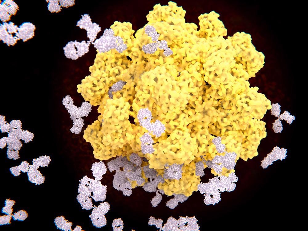
Immunoprecipitation (IP) is one of the commonly used techniques based on antibody-antigen interaction. This technique has various applications, including the isolation of proteins from biological samples. Further studies on these proteins are essential for their identification, structural characterization, and elucidating post-translational modifications.
IP is a precipitation technique that is commonly used for the isolation of proteins and other biomolecules from cell or tissue lysates. Cell lysates are comprised of mixtures of proteins, lipids, carbohydrates, and nucleic acids. Researchers use specific capture antibodies or protein beads that can bind to the target molecules. Subsequently, the targeted protein is separated and subjected to further studies using immunoblotting assays.

Image Credit: Juan Gaertner/Shutterstock.com
Factors Associated with Immunoprecipitation Technique
IP is based on the affinity between an antibody and its target protein. While conducting IP analysis, the antibody is incubated with lysates that contain the target protein. During the incubation period, an antibody-protein complex is formed, which is subsequently captured by antibody-binding proteins attached to agarose or magnetic beads. The target protein can then be eluted from the beads or agarose for further analysis.
While performing IP experiments, researchers have to consider some factors to obtain better results. Firstly, while preparing cell lysates, it is important to use the right lysis buffer (e.g., NP-40, Triton X-100) to ensure high-quality samples for IP. Non-specific binding between antibodies and proteins in the cell lysates could be reduced by incubating with protein beads and/or isotype control antibodies before performing IP. This step is mainly used while conducting co-immunoprecipitation (Co-IP).
Another important aspect of IP assay is the selection of antibodies. Capture and detection antibodies must be selected such that they originate from different species (e.g., rabbit capture antibody and mouse detection antibody). This is important to prevent interference from the heavy and light chains of the capture antibody during the detection process.
In IP assay, agarose and magnetic beads are commonly used supports. Agarose beads have a high antibody binding capacity due to their sponge-like structures and require longer incubation with the antibody-protein complex.
On the other hand, magnetic beads are much smaller than agarose beads with smooth outer surfaces. It has a relatively lower binding capacity compared to agarose beads. One of the advantages of using magnetic beads is a shorter incubation time due to a higher diffusion rate.
Different Types of Immunoprecipitation (IP) Assays
Different types of IP assays are used to study the interaction between the unknown (target) protein and other proteins or nucleic acids. Some of these types are discussed below:
Individual Protein Immunoprecipitation (IP): Individual Protein IP utilizes an antibody to isolate a specific protein of interest from cell lysate. The antibody binds to the protein and subsequently, the antibody-antigen complex is extracted using protein A-G-coupled agarose or magnetic beads.
Next, these beads are washed and the protein is eluted. The purified antigen, obtained by IP, is analyzed using varied molecular techniques such as ELISA and Western blot. The extracted protein is detected and quantified via mass spectrometry using enzymatic digestion patterns based on the primary sequence.
Co-Immunoprecipitation: This assay is also known as Co-IP. In this type of immunoprecipitation assay, researchers can pull down multiple proteins along with the target protein as a complex. For example, researchers can pull down clathrin-binding proteins that would help in the understanding of how these proteins enter or exit cells.
Therefore, this technique can map out proteins' interactions with each other, leading to particular functional phenotypic effects. Typically, Co-IP is only used to determine protein interactions between suspected interaction partners. This technique is not suitable for the high-throughput screening approach.
Chromatin Immunoprecipitation: This immunoprecipitation technique is also known as ChIP. In this method, the target protein is firstly bound with DNA and, subsequently, cross-linked by using formaldehyde to lyse the cells. The DNA is fragmented such that the target protein is only bound to the DNA that it interacts with.
After precipitation of protein occurs, it can be easily separated from the DNA using heat. Next, techniques such as Polymerase Chain Reaction (PCR) could be used to determine the specific DNA fragments it is attached to.
RNA Immunoprecipitation: This type of immunoprecipitation is also known as RIP which targets RNA-binding proteins (ribonucleoproteins). Similar to ChIP, RIP pulls out proteins that bind to RNA inside cells, and the RT-PCR technique is used to analyze the RNA obtained.
Advantages and Applications of Immunoprecipitation
Two of the main advantages of the IP technique are its speed and simplicity of the analysis in comparison to affinity chromatography, which is extremely time-consuming as it involves multiple cycles of binding and washing. Among the different types of immunoprecipitation assays, Co-IP is relatively simple and compatible with many protein analysis processes, such as western blog and mass spectrometry. Co-IP is used to identify protein interactions with physiological importance.
Originally, IP was developed as an alternative technique to column affinity chromatography for preparing a small number of protein samples. This sample was subsequently analyzed using mass spectrometry, western blot, etc. IP can be used to detect and identify proteins, present in minimal quantities, from various biological sources.
Researchers have revealed that IP is the main technique in post-translational modification detection assays. Co-IP helps identify physiologically relevant protein interactions. It is also used to capture low affinity and transient protein interactions. This technique can also detect the protein-protein interactions indirectly where a third protein could be sandwiched in between the bait and target proteins.
In summary, the main applications of IP are isolation and detection of proteins of interest, enrichment of low abundant proteins, and gaining a better understanding of the protein-protein interactions. Additionally, IP helps to detect unknown proteins in a protein complex and corroborate the expression of a protein in a specific tissue.
Sources:
- Shi, G. (2021) Immunoprecipitation (IP) Principles and Factors to Consider to Obtain Quality Results. [Online] Available at: https://blog.benchsci.com/immunoprecipitation-ip-principles-and-troubleshoot
- Overview of the Immunoprecipitation (IP) Technique.Thermo Fisher. (2021) [Online] Available at: https://www.thermofisher.com/uk/en/home/life-science/protein-biology/protein-biology-learning-center/protein-biology-resource-library/pierce-protein-methods/immunoprecipitation-ip.html
- Corthell, t.J. (2014) Immunoprecipitation. Basic Molecular Protocols in Neuroscience: Tips, Tricks, and Pitfalls, Academic press. pp. 77-81. https://doi.org/10.1016/B978-0-12-801461-5.00008-3.
- Kaboord, B. and Perr, M. (2008) Isolation of proteins and protein complexes by immunoprecipitation. Methods Molecular Biology. 424, pp. 349-64. doi: 10.1007/978-1-60327-064-9_27.
Further Reading