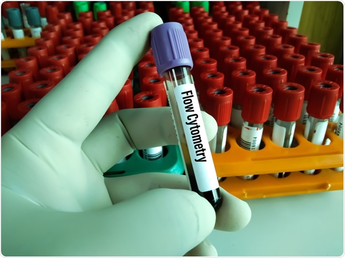
Immunophenotyping is used for antigen and surface marker recognition
Immunophenotyping is a process used to identify cells based on the antigens or markers expressed on the cell surface. Two main methods are used, flow cytometry immunophenotyping (FCI) and immunohistochemistry (IHC). FCI is a light-based analysis procedure capable of rapidly analyzing and quantifying cells in suspension that have been labeled with fluorescent antibodies. IHC is the fluorescent immunolabeling of cells in culture to observe morphology and distribution which can’t be observed with FCI. Both techniques are incredibly effective but vary in sensitivity depending on their application.
Several different types of tissue can be analyzed by FCI or IHC, but their diagnostic value is seen clearly in blood cancers, such as leukemia. FCI is the preferable approach in diagnostics due to its speed and ease, but both processes can be done with relatively little sample volume.
A method developed as an alternative to FCI is chip cytometry, a slide-based cytometry like IHC, however, it uses a staining method that could theoretically allow an unlimited number of analyses on one sample. Currently, chip cytometry has only been applied to peripheral blood mononuclear cells and bronchoalveolar lavage cells.

Image Credit: Arif biswas/Shutterstock.com
FCI and IHC may need to be used in tandem for accurate diagnoses
FCI gives detailed analyses of cell populations, providing information on more than one specific antigen per test and is sensitive to weakly expressed antigens. Additionally, sample acquisition is less invasive than a traditional biopsy necessary for IHC. Issues arise with the usage of bone marrow samples which may provide sclerotic or dense samples wherein there are too few adequate cells for analysis.
While also well used in hematology, issues arise in diagnosing lymphomas where B-cells cannot be identified in T-cell rich lymphoma subtypes, inability to detect malignant T-cells without aberrant immunophenotypes, and inability to detect Hodgkin lymphoma due to the low numbers of neoplasms.
Despite these specific shortcomings, FCI is effective as a first-line diagnostic method in patients with suspected hematological abnormalities. In the absence of meaningful results, it is recommended to be correlated with light microscopy for histological analysis and IHC in some cases. IHC is superior in its ability to detect relatively low numbers of neoplastic cells, such as that in Hodgkin lymphoma, but it is arguably impossible to detect weakly expressed antigens in paraffin tissue.
When fixing a sample for IHC, it is understood that the fixation process may compromise the tissue integrity, especially compared to FCI. Additionally, some antibody markers are simply not routinely available for paraffin IHC, such as CD13 and CD14, but can be used in FCI.
Primarily, FCI is the preferred method of immunophenotypically differentiating leukemia subtypes. It assists by more objectively confirming the presence of hematopoietic stem cells which provides the specificity for the diagnosis of acute leukemia. As well as diagnosis, FCI can monitor and assess residual disease markers and give an insight into the state of patient prognosis and remission.
In some cases of acute lymphoma, FCI can detect differences in levels of the antigen HLA-DR, the levels of which can indicate whether a patient is standard, medium, or high-risk which can influence the stage and therefore the treatment protocol in the patient. Overall, it seems that FCI can be sufficiently powerful on its own for certain diagnostic cases but requires additional validation from IHC or similar histological analyses to ensure accuracy or to identify diagnoses that would otherwise have been missed.
Cerebrospinal fluid contents can be immunophenotyped using chipcytometry
Cerebrospinal fluid (CSF) is rich in immunological markers useful for the diagnosis of a variety of different disorders. The gold standard of CSF immunophenotyping is FCI, but the emergence of chip cytometry as a method of immunophenotypic analysis shows promise for both research and diagnostic purposes.
FCI typically elucidates the pathophysiology of neuroinflammatory disorders such as multiple sclerosis (MS) wherein the immune cell composition of the CSF, as in leukemia, can be particularly valuable indicators of prognosis and assist treatment decisions. However, FCI is rarely used as the procedure for obtaining CSF via lumbar puncture is invasive and perceived as painful by many patients. For this reason, it is performed sparingly on patients and rarely performed repetitively.
FCI cannot be performed repeatedly on the same sample throughout the diagnostic and monitoring process if more questions arise, FCI relies on identifying pre-determined antigen markers which may change or evolve as more is elucidated from the patient's symptoms. Chipcytometry is based on microfluidic chips containing cell-adhesive surfaces allowing for quick and easy slide preparation, repeated staining, and long-term storage.
Repeated staining is facilitated by iterative staining-imaging-bleaching cycles which could theoretically allow an unlimited number of analyses for various cell surface antigens. To assess the viability of chip cytometry on CSF analysis, a study used CSF samples from over 350 patients with a variety of conditions.
Results show that low cell density (<5/μl) hampers the accuracy of the analysis and makes the procedure more time-consuming. The scanning procedure of the microscope used is also time-consuming; cell recognition becomes more error-prone when fewer cells are available, this can be attributed to the comparatively low leukocyte count in CSF in comparison to the peripheral blood for which this method was designed.
Additionally, cell loss was reported due to periods of washing which seems to be unique to CSF cells as similar results were not seen in studies using cells of different sources. Despite this, the results from chipcytometer use are impressive with slides retaining biomarker stability up to 20 months after sample preparation with the ability to establish additional markers at any time point.
Immunophenotyping is a valuable tool for diagnosis and research
Immunophenotyping is a powerful tool, particularly in differentiating between subtypes of similar diseases. The two main methods, FCI and IHC, have their benefits and drawbacks but there is room for improvement in the field which is quickly being filled with developments such as chip cytometry.
Sources:
- National Cancer Institute. (2021). Immunophenotyping. [Online] National Cancer Institute. Available at: www.cancer.gov/.../immunophenotyping (Accessed on 1 September 2021)
- Brown, M., et al. (2000). Flow Cytometry: Principles and Clinical Applications in Hematology. Clinical Chemistry. https://doi.org/10.1093/clinchem/46.8.1221
- Abdel-Ghafar, AA., et al. (2012). Immunophenotyping of chronic B-cell neoplasms: flow cytometry versus immunohistochemistry. Hematology Reports. https://doi.org/10.4081/hr.2012.e3
- Dunphy, CH. (2004). Applications of Flow Cytometry and Immunohistochemistry to Diagnostic Hematopathology. Archives of Pathology & Laboratory Medicine. https://doi.org/10.5858/2004-128-1004-AOFCAI
- Wood, BL. (2014). Flow cytometry in the diagnosis and monitoring of acute leukemia in children. Journal of Hematopathology. https://doi.org/10.1007/s12308-014-0226-z
- Ouyang, G., et al. (2019). Clinically useful flow cytometry approach to identify immunophenotype in acute leukemia. Journal of International Medical Research. https://doi.org/10.1177/0300060518819637
- Hümmert, MW., et al. (2018). Immunophenotyping of cerebrospinal fluid cells by Chipcytometry. Journal of Neuroinflammation. https://doi.org/10.1186/s12974-018-1176-7
Further Reading
Last Updated: Nov 12, 2021