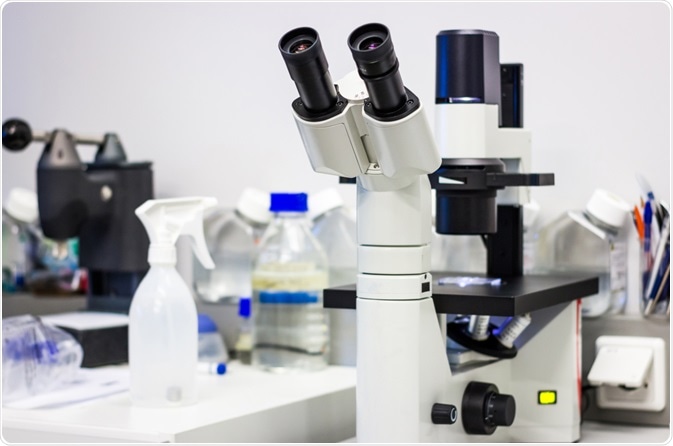
Phase-contrast microscopy is a contrast-enhancing technique used to image transparent specimens. A Dutch physicist, Frits Zernike first described phase-contrast microscopy and was awarded a Nobel Prize in Physics in 1953 for his discovery.

Image Credit: Catalin Rusnac/Shutterstock.com
This technique uses the minute variations in-phase and the optical path length within the specimen to enhance contrast and visualize transparent materials. Some of the transparent specimens that can be viewed using phase-contrast microscopy include microorganisms, living cells, tissue sections, and other materials such as fibers, glass fragments, etc.
Background
Light travels in waves (sine waves) consisting of repeated crests and troughs. The human eye perceives the intensity or brightness of the light based on the amplitude or height of the wave. The color of the light is determined by the wavelength, which is the distance between two successive crests or troughs of a wave.
The phase of a wave refers to where along the sine wave the wave is at a given point of time. For example, the wave could be at the peak of the sine wave or the bottom of the trough, or in between depending on a given point of time.
Light travels at a speed of 300,000 km/sec in a vacuum. However, this speed varies when it passes through a denser medium as compared to a less dense medium. The degree to which the light speed varies depends on the refractive index of the medium. Therefore, light travels slower through thicker specimens with a high refractive index and vice versa. The distance light has to travel through a medium is known as optical path length.
Transparent specimens such as cells do not absorb light and are called phase objects. When light passes a part of the specimen, such as a sub-cellular structure, it results in a small change in refractive index and optical path length.
Working model of phase contrast microscopes
The partially coherent light emitted from an illumination source such as a tungsten-halogen lamp will be passed through the collector lens and then focused on the condenser annulus positioned in the substage condenser front focal plane. The specimen will then be illuminated by the wavefronts passing through them.
Depending on the object in the light path coming from the condenser annulus, the wavefronts can either pass through without being deviated or diffracted and retarded in phase by structures and phase gradients present across the specimen. The un-deviated and diffracted wavefronts will then be collected by the objective and segregated by a phase plate at the rear focal plane and focused on the intermediate image plane to form the final phase-contrast image.
Brightfield microscopy Vs phase contrast microscopy
Visualization of unstained biological specimens such as cells produces low contrast images because these specimens don’t absorb light due to their high-water content. To overcome this challenge, biological samples are often stained with light-absorbing dyes. Therefore, images obtained from brightfield illumination of stained specimens rely on the absorption of the visible light by the staining dyes.
However, staining the cells will not be appropriate for some applications. In such cases, a phase-contrast microscope would be well suited as it provides high contrast images of transparent specimens. By relying on the changes in the refractive indices of the different components or optical path lengths, specimens can be imaged in their native unaltered state using phase-contrast microscopy.
Advantages of phase contrast microscopy
One of the major advantages of using a phase-contrast microscope is the ability to image live cells without altering them in any way. Traditional brightfield microscopy techniques for imaging transparent sections would include several steps such as fixation, tissue processing for histology, and cutting and tissue staining with dyes to differentiate the cellular structures.
Each of these steps may alter the physiological state of a biological specimen. On the other hand, phase-contrast microscopy does not rely on these steps, therefore, the cells could be examined in their native state. A phase-contrast microscope equipped with modern accessories can perform live-cell imaging as well as time-lapse of a particular specimen by video recording.
Advancements in phase-contrast microscopy
The addition of phase contrast optical accessories to a standard brightfield microscope can enhance contrast to the transparent specimens. The current generation phase contrast microscopes are equipped with imaging modalities coupled with excellent electronic enhancement and post-processing image enhancing techniques. These advancements allow the users to image specimens with excellent contrast between subcellular structures or very small internal structures.
Further Reading