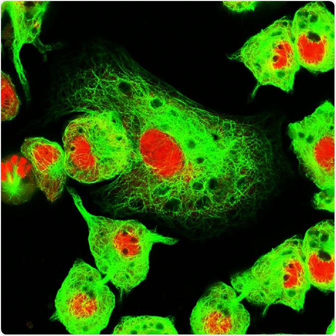
Photobleaching is the phenomenon when a fluorophore loses its fluorescence due to damage induced by light. This leads to loss of fluorescence and signal while imaging a sample.

Real fluorescence microscopic view of human neuroblastoma cells. Image Credit: Vshivkova / Shutterstock.com
The Principle
When the light of appropriate wavelength is directed on a fluorophore, it transitions from the ground state to excited singlet and triplet stages. In its excited state, it may interact with other molecules and undergo permanent covalent modifications.
Also, the triplet state is long-lived in comparison to the singlet stage, which makes it more susceptible to interactions with other molecules. The total number of these cycles between excitation and emission for each fluorescent molecule is dependent on its structure and the local environment around it.
Based on these parameters, some fluorophores can bleach quickly, while others can undergo several thousands of cycles of excitation and emission before bleaching.
Methods to Reduce Photobleaching
Reducing the intensity of light
Reducing the intensity of light can reduce the exposure of the fluorophores to lights, and thus, reduce the excitation-emission cycles it undergoes. This eventually reduces the photobleaching associated with it. However, reducing the light intensity can also reduce the intensity of the signal. Therefore, a correct balance should be made between achieving the right signal intensity and minimal photobleaching.
Reducing the time exposure to light
This is another method to reduce the photobleaching effect. In this case, reducing the time of exposure to light will reduce the number of times the fluorophores undergo the excitation-emission cycles, thus reducing the photobleaching. This can be achieved by reducing the pixel dwell of laser light or fixing a faster frames/sec rate of imaging.
Another method is to limit the region of imaging. This can be achieved by fixing a small region of interest, and after imaging that area, moving on to the adjacent area.
Addition of antifade
Antifades are commercially available reagents that can reduce the photobleaching effect. These are mounting media, which are used after all the steps of immunocytochemistry in the final step before imaging.
Different antifade reagents are available that can be tested to see which reagent has the optimal effect for your sample.
Using a different dye
Different dyes or fluorophores have different photobleaching properties. For example, red fluorescent proteins are more susceptible to photobleaching compared to green fluorescent proteins. Thus, changing the chosen dye and using one that is more photostable can also be employed to reduce photobleaching of the sample.
Using neutral-density filters
The use of neutral density filters can also reduce the light or photon numbers incident on the sample. This can also reduce the exposure of the sample towards light and reduce photobleaching. Again, this can also reduce the signal of the sample.
One method to overcome this is by increasing the gain function of the photomultiplier. However, increasing this function also increases the noise along with the image signals. Thus, an appropriate balance between signal-to-noise ratio and photobleaching should be achieved to get the best image.
Productive Uses of Photobleaching
Autofluorescence is the phenomenon when the sample contains cellular objects that are naturally fluorescent due to endogenous fluorophores. These tissues or organelles often fluoresce at similar wavelengths as commonly used fluorophores, such as green fluorescent protein (GFP) or fluorescein isothiocyanate (FITC).
This increases the error in distinguishing between the actual signal and the signal from endogenous fluorophores. In such cases, photobleaching can be used as a tool.
The image can be exposed to UV rays for two hours in order to bleach the endogenous fluorophores and then subsequently imaging the sample with appropriate light specific fluorescent proteins, such as GFP, to reduce the autofluorescence effect.
Further Reading
Last Updated: Feb 1, 2021