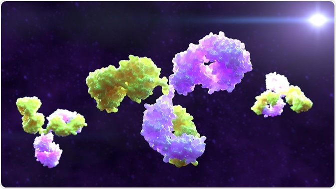
What is monoclonality and why it is important to study it?
Monoclonality is a term used in biology and medicine referring to a monoclonal cell or a substance. To be defined as monoclonal the cell or substance has to have been derived from a single clone, whereas if it is polyclonal it has been derived from many clones.

Image Credit: Alpha Tauri 3D Graphics/Shutterstock.com
Often, monoclonality is studied in oncology to determine the origin of a malignant tumor. Usually, in the early stages of tumorigenesis, the cancerous cells are monoclonal. Later on, as the tumor progresses, these monoclonal cells may acquire a mutant phenotype leading to the formation of subclones.
Clonal overgrowths can indicate neoplastic proliferation and their clinical detection has been used for several years to diagnose illnesses such as malignant lymphomas. The technique is being developed for use in many applications in tumor pathology, which may help to diagnose, monitor, and improve treatment outcomes for some types of cancer.
What is PCR and how is it used to detect monoclonality?
Polymerase chain reaction (PCR) is a method used to rapidly make numerous copies of a specific DNA section of clinical or research interest. The basic aim of PCR is to replicate enough of the section of desired DNA so that there is a sufficient amount to run an analysis on it, or for it to be used in some alternative way.
The technique relies on a thermostable DNA polymerase and uses specially created DNA primers that are suited to the target segment of DNA. The PCR reaction is replicated, cycling through multiple changes in temperature so that numerous copies of the selected DNA can be generated.
While PCR has many firmly established uses in forensic analysis and DNA cloning, its capability of detecting monoclonality has contributed to its application in medical diagnostics.
As clonal overgrowths are indicative of tumor proliferation, their identification can be used to diagnose cancers such as malignant lymphomas. To demonstrate clonal overgrowth, scientists can rely on the detection of specific dominant immunoglobulins and/or T-cell receptors.
Also, nonrandom genetic alterations can be used to evaluate the clonal expansion and clonal evolution of tumors, analyzing hypervariable segments of DNA in patients who express heterozygous alleles for a specific marker.
These tests are based on the recognition of a loss of heterozygosity (LOH) due to nonrandom interstitial DNA deletions or resulting from the homozygosity of mutated alleles that occurs in neoplasms. These LOH tests highlight the clonal expansions of tumor cells and identify monoclonal proliferation in cases when multiple and consistent LOH is observed.
PCR can be used to prove monoclonality in a couple of ways. The first is through the analysis of microsatellites (linked or not to X chromosome). The second is through gene rearrangement detection.
Microsatellites
Microsatellites refer to short, repeated sections of DNA that occur at a particular locus on a chromosome. They are not uniform across the population and therefore can be used for genetic fingerprinting. X chromosome related markers can provide clonality information using LOH analyses following DNA digestion with methylation-sensitive endonucleases.
Non-X chromosome related markers can be used in conjunction with this information to provide complementary data. In this way, researchers can obtain information that is both related and not related to the transformation pathway of the tumor.
When used appropriately, these tools can demonstrate monoclonality and determine the clonal evolution of a tumor cell population, enabling scientists to distinguish recurring tumors from other metachronic neoplasms, flag early relapses, and discriminate field transformation from metastatic tumor growths.
Gene rearrangements
PCR can also be used to prove monoclonality by analyzing gene rearrangements. This technique has proven useful in the diagnosis of malignant lymphomas. Malignant lymphomas go through certain genetic mutations involving the processes of deletion, nucleotide additions, and specific splicing.
The PCR design for gene rearrangement detection takes these DNA sequence additions and deletions into consideration.
In this circumstance, the primer binding regions chosen for the PCR process should be located within less variable sequences of DNA so that failing amplification due to the loss of these regions can be avoided. False-negative results in detecting clonal rearrangements can occur if the primer locations are not suitable. Also, these PCR analyses require careful optimization and suitable controls, such as internal positive, positive, negative controls to again minimize the risk of generating false results.
Using this system, well-defined monoclonal PCR bands can be detected even when trace amounts of lymphoid DNA are present.
Summary
PCR is a reliable method of proving monoclonality. It has become firmly established as a tool used to assist in the diagnosis of various cancers. This technique can be used to demonstrate monoclonality through the analysis of microsatellites as well as by looking at gene rearrangements.
Both these approaches have specific setups, drawbacks, and uses, and both have become well-established in the monitoring and diagnosis of malignant lymphomas.
Sources:
- Cozzio, A. and French, L. (2008). T-Cell Clonality Assays: How Do They Compare?. Journal of Investigative Dermatology, 128(4), pp.771-773. https://www.sciencedirect.com/science/article/pii/S0022202X15338057
- Diaz–Cano, S., Blanes, A. and Wolfe, H. (2001). PCR Techniques for Clonality Assays. Diagnostic Molecular Pathology, 10(1), pp.24-33. https://www.ncbi.nlm.nih.gov/pubmed/11277392
- Weirich, G., Funk, A., Hoepner, I., Heider, U., Noll, S., Ptz, B., Fellbaum, C. and Hfler, H. (1995). PCR-based assays for the detection of monoclonality in non-Hodgkin's lymphoma: application to formalin-fixed, paraffin-embedded tissue and decalcified bone marrow samples. Journal of Molecular Medicine, 73(5). https://www.ncbi.nlm.nih.gov/pubmed/7670927
Further Reading
Last Updated: Mar 18, 2020