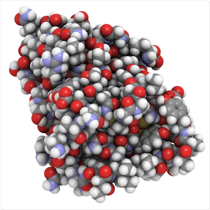
What are nanobodies and why are they important?
A nanobody is a small antibody fragment consisting of a single monomeric variable antibody domain. Nanobodies were first engineered from heavy-chain antibodies found in camelids (camels, llamas, alpacas, and dromedaries).

Image Credit: StudioMolekuul/Shutterstock.com
These nanobodies have special properties that make them useful in a variety of scientific applications. Studies have shown that nanobodies have the full antigen-binding capacity and that they are also incredibly stable since they can maintain the unique functional and structural properties of the heavy chain antibodies, from which they are derived.
In addition, nanobodies are the smallest naturally occurring integral antigen-binding units, therefore they are particularly useful as components of novel biological drugs.
These special properties have made nanobodies vital to the development and large-scale production of a wide range of innovative biological therapeutics that are being established to tackle several human diseases.
Previously, monoclonal antibodies that are generally derived from mouse hybridomas have been used in various diagnostic and therapeutic tools due to their efficiency and antibody-antigen specificity.
However, there are several drawbacks related to the use of monoclonal antibodies. For instance, they are relatively expensive to use, and their large size limits accessibility and tissue penetration.
In recent years, genetic engineering techniques have rapidly advanced. As a result, some antibody fragments have been developed into viable alternatives to monoclonal antibodies, such as nanobodies.
Several methods are currently available to construct a nanobody library. Currently, the most popular method is phage display, where libraries are generated from immunized camelids.
However, this method has limitations in that while high-affinity nanobodies can be isolated rapidly, in some cases the antigen of interest is toxic to the camel making immunization impossible, or the antigens are produced as inclusion bodies with wrong folded proteins.
To overcome these problems, synthetic phage display libraries generated with the polymerase chain reaction (PCR) have been developed as an alternative method.
Creating a library with the help of PCR
Several years ago, scientists established a reliable method for creating nanobody libraries based on synthetic phage display techniques.
A team of researchers based in Belgium created a synthetic phage display nanobody library using the conserved camel single-domain antibody fragment (VHH) framework of cAbBCII10. Below, we discuss the method that they developed, and explain the role of PCR in this approach.
In creating this library, the team followed the universal VHH structure of cAbBCII10 that was outlined previously by some of the same scientists in another research group.
The team first introduced diversity in the complementarity-determining region 3 (CDR3) by randomization of synthetic oligonucleotide DNAs using the codon NNK for the library DNA construction. The primers for the PCR were generated using a specific DNA sequence of the cAbBCII10 structure.
Next, the synthetic nucleotides were amplified by overlap extension PCR to create numerous copies. The restriction enzymes Pst I and Not I were used to digest the resultant PCR products, which were then separated via agarose gel electrophoresis and extracted. Next, the PCR fragments were cloned into the phagemid vector pMECS to multiply them, and the clones were used to transform Escherichia coli (E. coli) TG1 cells by electroporation.
The clones were left overnight at 28°C on plates in growth medium containing 2× yeast-extract-tryptone, ampicillin, and glucose. Researchers were able to confirm the size of the library by summating the colonies present following a gradient dilution.
At the same time, the team randomly selected 24 clones to perform additional PCR to establish the correct rate of recombinant DNA insertion. Also, 30 more clones were randomly selected and sequenced to uncover the amino acid composition and distribution of CDR3.
The final step saw the colonies removed from their plates, added to a medium containing 1% glucose and 50% glycerol, and stored at −80°C until needed.
An effective method established
The protocol established by the Belgian researchers was proven successful for the construction of a large and diverse synthetic phage display nanobody library based on the conserved camel single-domain antibody fragment (VHH) framework of cAbBCII10 that members of the team had identified in a previous project. Results showed that diversity was successfully introduced in the complementarity-determining region 3 (CDR3) through the process of synthetic oligonucleotide randomization.
Nanobodies from libraries generated using this method have been selected by other research groups through the use of specific antigens such as human pre-albumin (PA) and neutrophil gelatinase-associated lipocalin (NGAL).
Conclusion
The researchers proved that the protocol they established works as a reliable source for producing antigen-specific nanobodies. They believe that the approach will likely be useful in providing the medical community, particularly the field of medical diagnostics, with a resource of nanobodies for the development of diagnostic methods.
Sources:
- Pardon, E., Laeremans, T., Triest, S., Rasmussen, S., Wohlkönig, A., Ruf, A., Muyldermans, S., Hol, W., Kobilka, B. and Steyaert, J. (2014). A general protocol for the generation of Nanobodies for structural biology. Nature Protocols, 9(3), pp.674-693. https://www.ncbi.nlm.nih.gov/pmc/articles/PMC4297639/#!po=82.5000
- Saerens, D., Pellis, M., Loris, R., Pardon, E., Dumoulin, M., Matagne, A., Wyns, L., Muyldermans, S. and Conrath, K. (2005). Identification of a Universal VHH Framework to Graft Non-canonical Antigen-binding Loops of Camel single-domain Antibodies. Journal of Molecular Biology, 352(3), pp.597-607. https://www.ncbi.nlm.nih.gov/pubmed/16095608
- Yan, J., Li, G., Hu, Y., Ou, W. and Wan, Y. (2014). Construction of a synthetic phage-displayed Nanobody library with CDR3 regions randomized by trinucleotide cassettes for diagnostic applications. Journal of Translational Medicine, 12(1). https://www.ncbi.nlm.nih.gov/pmc/articles/PMC4269866/#!po=68.3333
Further Reading