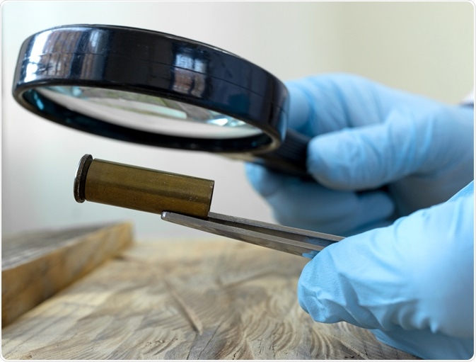
Ballistic trauma is extremely significant to criminal investigations involving the use of firearms; however, much of the research in this field has been limited to macroscopic evaluation of evidence. To improve the precise diagnosis and evaluation of ballistic trauma, several efforts have been made to increase the use of microscopy for this purpose.

Ballistics. Image Credit: Sendo Serra/Shutterstock.com
An introduction to forensic ballistics
By definition, forensic ballistics investigates the motion of the projectile from the moment that ammunition is released by the firearm until it comes to a rest. Forensic ballistics is therefore notably different than forensic firearm examination, which involves the examination of firearm evidence that has been collected from a crime scene to determine their forensic value and role in the crime.
Forensic ballistics can be further subdivided into three distinct areas, which include internal, external, and terminal ballistics. Internal ballistics refers to all movement experienced by the projectile before it is released from the firearm, whereas external ballistics refers to all motion of the projectile from the moment it is released by the firearm and as it travels through the atmosphere.
Finally, terminal ballistics describes the motion of the projectile as it comes into any type of physical matter. An additional branch of forensic ballistics includes intermediate ballistics, which is used to describe the motion of the projectile as it moves away from the barrel of the firearm until escapes the flow of gases and enters free light.
The utility of microscopy in ballistics
Small firearms are implicated in up to 40% of homicides, 6% of suicides, and up to 90% of all conflict deaths in the human population. Therefore, the accurate interpretation of ballistic trauma is crucial in establishing the circumstances that surrounded the injury or death, particularly in criminal cases.
Despite the utility of microscopic investigations in ballistic trauma, particularly those involving skeletal bones, macroscopic examination has often been the method of choice for forensic investigators. This preference for macroscopic analysis has largely been due to the assumption that the unique morphology of ballistic skeletal trauma can be diagnosed with the naked eye.
Improving beveling inconsistencies
One of the most advantageous applications of microscopy in forensic ballistics, as opposed to macroscopic investigation, can be found in the beveling of entry and exit wounds. The beveling of skeletal bone, which indicates that there has been a change in the square edge of the bone to one that is now sloping, is almost always indicative of bullet trauma. Despite this association, beveling is often absent when thinner bones such as the sphenoid, orbit, or squamous temporal bone, as well as juvenile bones, are analyzed.
In addition to its lack of macroscopic specificity on thin bones, beveling may not always indicate bullet trauma. For example, one study found that a circular cranial defect with internal beveling that was originally diagnosed as a gunshot trauma following macroscopic evaluation was subsequently found to be the result of blunt trauma once the bone sample was analyzed by scanning electron microscopy coupled with dispersive X-ray spectrometry (SEM-EDS).
SEM in forensic ballistics
As a result of the deficiencies associated with the macroscopic analysis of ballistic trauma in bones, it is currently recommended that optical microscopic analysis is used to evaluate bullet wipes and link bony defects to the projectile that caused them. In addition to optical microscopy, several studies have been published evaluating the utility of SEM for the finer analysis of ballistic trauma.
In 2014, an in-depth study was conducted to ultimately determine how SEM could improve forensic ballistic diagnoses. This study found that SEM could successfully provide directionality signatures that could be used to determine both the entry and exit status in bone architecture after projectile insult.
Additionally, this study found that although thin bones may not allow for bevel formation to occur, SEM can be used to analyze deflections of even small broken bone fragments to determine the trajectory. Previously, thin bones with ballistic trauma that do not present with beveling would leave forensic investigators unable to accurately determine entry or exit status. This discovery, therefore, puts SEM in a unique position to resolve some of the traditional issues that forensic investigators encounter when looking to determine shot direction in these bone samples.
SEM has also been found to assist in distinguishing between perimortem from postmortem lesions. A perimortem lesion within a bone will often retain some of its organic constituents, which is comparable to postmortem lesions that will instead be lost upon SEM analysis of the fracture morphology.
A perimortem lesion can also be determined upon the identification of any fibrous connections, which are high in organic content, that remain between the fractured lamellae of bones. Since bones may retain organic content for extended periods, the absence of certain defects upon SEM analysis such as cortical strands and/or bending deformations cannot definitively rule out that the lesion is of postmortem origin.
Sources:
- Ballistics [Online]. Available from: https://www.nist.gov/ballistics.
- Bolton-King, R. S. (2016). Preventing miscarriages of justice: A review of forensic firearm identification. Science & Justice 56(2); 129-142. doi:10.1016/j.scijus.2015.11.002.
- Rickman, J. M., & Smith, M. J. (2014). Scanning Electron Microscope Analysis of Gunshot Defects to Bone: An Underutilized Source of Information on Ballistic Trauma. Journal of Forensic Sciences 59(6); 1473-1486. doi:10.1111/1556-4029.12522.
- Vorburger, T. V., Song, J., & Petraco, N. (2016). Topography measurements and applications in ballistics and tool mark identifications. Surface Topography 4(1). doi:10.1088/2051-672X/4/013002.
Further Reading
Last Updated: Mar 11, 2021