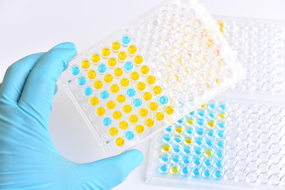
Immunoassays (IAs) are extremely important bioanalytical methods that are used to quantify analytes (blood serum, toxin, hormone, etc.) ranging from small molecules to macromolecules, in a solution, using antibody or antigen as a biorecognition agent. Immunoassays are applied in wide-ranging scientific disciplines that include biopharmaceutical analysis, clinical diagnostics, environmental monitoring, and food testing.

Image Credit: Jarun Ontakrai/Shutterstock.com
In 1917, Karl Landsteiner, an Austrian biologist, first developed methods for the analysis of small molecules with antigenic properties. His method became the framework for the development of immunoassays. Between 1995 and 2017, scientists have developed a broad range of immunoassays that enabled qualitative and quantitative detection of biological samples.
IAs play a crucial role in clinical decision-making, and are, therefore, indispensable for healthcare settings. Most of the immunoassay methods are based on radioimmunoassay (RIA) and enzyme immunoassays (EIA). A common type of EIA is enzyme-linked immunosorbent assay (ELISA) which has many applications. Both ELISA and RIA-kits are commercially available for the detection of a wide range of analytes. These assays are extremely specific and highly sensitive and, thereby, serve as the gold standard for a plethora of analytes (antigen). The main advantages of immunoassays include inexpensiveness, the capability of detection of low levels of molecules, accuracy, and broad applicability.
Principle of Immunoassay
In response to foreign substances or antigens, such as bacteria and viruses, all animals and human beings produce antibodies via the immune system. Antibodies are Y-shaped, antigen-specific proteins that bind to an antigen at a specific site. As stated above, immunoassays are based on antibody-antigen binding relationships to detect specific molecules in a solution.
Classification of Immunoassays
Immunoassays are classified in various ways and one of the methods of classification is based on labeled and non-labeled immune assays. Labeled immunoassays are further classified as heterogeneous and homogenous immunoassays. A heterogeneous immunoassay is also known as separation immunoassay. It is a multiple-step method that is involved with the separation of analyte-antibody complex before analysis.
On the other hand, homogeneous immunoassays do not require separation before the analysis. Labeled immunoassays are also classified as competitive or non-competitive assays. In competitive immunoassays, the analyte (non-labeled antigen) competes with a specific amount of labeled antigen for the antibody, while non-competitive immunoassays use a surplus of labeled antibodies to bind with the analyte.
See how a lateral flow immunoassay works
Different Types Immunoassays Used in Scientific Research
Scientists have developed different types of immunoassays with wide-ranging applications. Some of the common types of immunoassays are discussed below:
Radioimmunoassay (RIA)
The radioimmunoassay method uses antibodies to quantify the amount of antigen present in a biological sample. This assay is extremely sensitive and specific, such that it can detect as low as a few pictograms of antigen in a sample. The basic principle of RIA is competitive binding. In this method, the target antigen is labeled using radioactive isotopes and bound to its specific antibodies.
The radioactive antigen competes with a non-radioactive antigen (from serum sample) for a fixed number of receptor binding sites or antibodies. The competition for the antibodies results in the release of a certain number of labeled antigens, such that it is proportional to the ratio of labeled to unlabeled antigen.
On increasing the concentration of unlabeled antigen, they displace bound labeled antigens. Subsequently, the bound antigens are separated from the unbound ones, and the radioactivity of the free antigens remaining in the supernatant is measured. The main advantage of the RIA method is the measurement of analytes with extreme precision and sensitivity.
Enzyme Immunoassay (EIA)
Although the main principle of enzyme immunoassay (EIA) is similar to RIA, it uses enzymes as the label instead of a radioisotope. In this assay, enzyme molecules are coupled into the immunoanalytical reagent via suitable reactions, and, subsequently, normal immunoassay procedures are performed.
After the separation of bound and free fractions, the enzyme activity is determined by adding a substrate. Owing to the enzymatic reaction a colored product is formed, which is measured using a spectrophotometer. The measured signal or the intensity of the color of the product is analogous to the concentration of the analyte.
Fluoroimmunoassay (FIA)
Fluoroimmunoassay is also similar to the working principle of RIA, except that the label is a fluorophore instead of a radioisotope. FIA is classified into heterogeneous and homogeneous assays, which are dependent on whether the separation step is required or not. These assays can be performed in a competitive or non-competitive format.
Heterogeneous competitive FIA methods are used to determine pharmaceuticals in biological samples, e.g., aminoglycoside antibiotics, morphine-3-glucuronide (the major urinary metabolite of heroin and morphine), benzoylecgonine in urine, etc. It is also applied for the determination of the total thyroxin in human serum.
The homogeneous competitive FIA method is used where the antibody binding leads to certain changes in the fluorescence properties of the labeled analyte. This method is also used to monitor the concentration of analyte directly from the reaction mixture.
Chemiluminescence Immunoassay (CLIA)
Chemiluminescence immunoassay is associated with chemiluminescent substance as a label. Typically, luminescence is the visible or near-visible radiation emitted when an electron transitions from an excited state to a ground state. This technique has been increasingly used in pharmaceutical analysis because of its low detection limits, high performance, and good specificity.
Liposome Immunoassay (LIA)
This immunoassay uses a liposome-encapsulating marker. Liposomes are used in LIA which are coupled with either analytes or antibodies and, thereafter, a normal immunoassay process is performed. Detection of LIA depends on the lysis of the liposome which releases the encapsulated marker. These measured markers correlate with the concentration of the analyte.
Capillary Electrophoresis Immunoassay (CEIA)
Capillary electrophoresis immunoassay is a relatively new technique that is applied for the sensitive detection of an analyte. This technique is based on the combination of the principles of capillary electrophoresis separation and heterogeneous immunoassay.
CEIA involves the covalent binding of an antibody to the modified interior surface of a microcapillary that serves as a solid phase immunoreactor. The detection of analytes is based on immunoassay. This method is extremely sensitive and is capable to detect analytes at picomole levels.
Sources:
- Gliechmann, N. (2020) Immunoassays: A Guide. Technology Networks. [Online] Available at: www.technologynetworks.com/.../immunoassays-a-guide-338790
- Vashist, K.S. and Luong, T.H.J. (2018) Immunoassays: An Overview. Handbook of Immunoassay Technologies, Academic Press. pp. 1-18.
- Cox, K.L. et al. (2012) Immunoassay Methods. [Online] Available at: https://www.ncbi.nlm.nih.gov/books/NBK92434/
- Darwish, I. A. (2006) Immunoassay Methods and their Applications in Pharmaceutical Analysis: Basic Methodology and Recent Advances. International Journal of Biomedical Science : IJBS. 2(3), pp. 217–235.
Further Reading