What is the post-mortem interval?
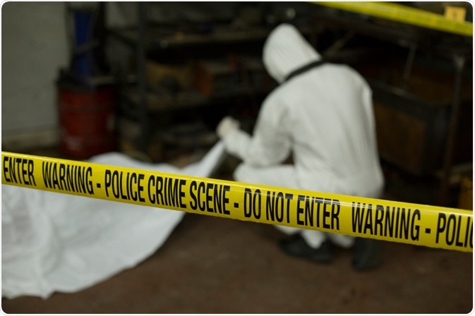
When a death occurs and no one is present to witness it, or in the case of a suspicious death the only witnesses may be the suspect(s) of the crime, the determination of post-mortem interval is vital to estimating when the death took place.

Image Credit: Wulle/Shutterstock.com
The post-mortem interval is determined as the length of time that has passed between the occurrence of death and the discovery of the body. It can also refer to the stage of decomposition the body is found in.
Determining the time of death can be incredibly valuable in helping investigators piece together the full story surrounding the death of an individual.
More importantly, it can help rule in or out certain possibilities, such as whether a particular suspect could potentially have been at the scene of the crime, or not, at the estimated time of death.
How is the post-mortem interval determined?
While determining the time of death is a crucial tool for forensic science, to date, a single precise method is yet to be established, and therefore a variety of methods are still used, such as body cooling, entomology, and putrefaction.
It is difficult to construct a method that can be fully relied on due to the variable nature of the changes that occur after death.
Various methods exist that analyze any other the number of changes that occur in the body’s tissues after death, however, the trajectory of these changes is often difficult to tie down to an exact time frame.
Since the post-mortem interval can be used as evidence in criminal cases, developing a reliable and accurate standardized measure of post-mortem interval is essential.
One such method that offers the potential to be utilized to determine the post-mortem interval is transmission electron microscopy (TEM). This is a powerful technique, estimating the post-mortem interval based on measurements of the dissolution of cells and tissues.
A recent study was conducted where this method was employed on gingival tissue obtained from deceased individuals with the goal being to monitor and record changes that occur in the tissue at intervals of two and four hours after death.
TEM revealed multiple ultrastructural changes in the gingival tissue. Two hours after death, lysis of the inner nuclear membrane was observed in the nuclei. Also, lysis of cristae was witnessed in mitochondria.
Lysis was also observed in the endoplasmic reticulum, which was also slightly swollen, and had released its ribosomes. Finally, an abundance of lysosomes was found in the intercellular areas.
After the passing of four hours since death researchers witnessed lysis of desmosome within the epithelium.
Also, the nucleus with nucleolus was recorded. Crenations in the nuclear membrane were observed in a few nuclei, and peripheral condensation of chromatin was also recorded.
The number of mitochondria had been reduced, and lysis of the outer membrane and the endoplasmic reticulum was observed. The level of the intracellular free ribosome was increased.
A comparative analysis of the changes witnessed in the tissue samples following two hours and four hours postmortem revealed that these morphological changes were directly dependent on the length of the post-mortem interval.
Therefore, these changes may be used to give an accurate and robust measurement of the post-mortem interval.
Currently, this method of PMI evaluation is in a developmental stage, with much more research needed to corroborate these findings and investigate cell changes at longer intervals before a reliable method can be established and made available to forensic crime labs.
However, so far, the results are promising, and it appears that TEM could complement the variety of methods currently used to measure the post-mortem interval.
Sources:
- Dix, J. and Graham, M. (2000). Time of death, decomposition, and identification. Boca Raton: CRC Press
- Muthukrishnan, S., Narasimhan, M., Paranthaman, S., Hari, T., Viswanathan, P. and Rajan, S. (2018). Estimation of time since death based on light microscopic, electron microscopic, and electrolyte analysis in the gingival tissue. Journal of Forensic Dental Sciences, 10(1), p.34. https://www.ncbi.nlm.nih.gov/pmc/articles/PMC6080157/
- Salam, H., Shaat, E., Aziz, M., MoneimSheta, A. and Hussein, H. (2012). Estimation of postmortem interval using thanatochemistry and postmortem changes. Alexandria Journal of Medicine, 48(4), pp.335-344. https://www.tandfonline.com/doi/full/10.1016/j.ajme.2012.05.004
- Madea, B. Methods for determining the time of death. Forensic Sci Med Pathol 12, 451–485 (2016). https://doi.org/10.1007/s12024-016-9776-y
Further Reading
Last Updated: Oct 5, 2020