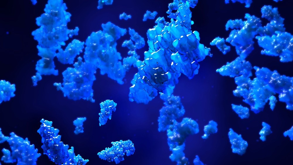
Proteomics involves all proteins produced by organisms (e.g., bacteria, plants, animals, humans) or living systems, including cell culture, organ, etc. Generally, two fundamental approaches are used to identify and characterize proteins, namely, the "bottom-up" and "top-down" methods. The present article focuses on "bottom-up" proteomics.

Image Credit: ArtemisDiana/Shutterstock.com
The field of proteomics progressed rapidly due to advancements in analytical tools, which are essential for studying proteins. For instance, the development of two-dimensional gel electrophoresis (2D-PAGE) has played an important part in the rapid separation of proteins from complex biological mixtures. Mass spectroscopy (MS) has played an important role in protein analysis among various analytical tools. This technique has helped accurately determine the mass and chemical structure of the proteins.
In a proteomic study, the continual large-scale production of peptide/protein sequences is stored in several databases. Scientists stated that bioinformatics tools play an important role in simplifying the analysis of a large volume of MS data for identifying and characterizing proteins.
Bottom-Up Proteomics: A Brief Overview
In most proteomic analyses, proteases are used to digest proteins into peptides. These peptides are then analyzed using an MS instrument, which provides information about the proteins present in the sample. Protein analysis converts proteins to peptides by digestion and subsequently characterizes the open reading frame of the peptides using protein databases. This analytical process has been grouped under "bottom-up" proteomics.
In short, a typical bottom-up proteomics process involves proteolytic digestion before analyzing it using MS. In bottom-up proteomics, the gel-based approach involves the separation of the protein mixture by electrophoresis, following which proteins are extracted from the gel and analyzed using MS. However, the gel-free approach involves direct digestion of protein mixtures, separation via multidimensional separation methods, and subsequent analysis of the resulting peptides using MS.
Sample Preparation and Mass Spectroscopic Analysis for Bottom-Up Proteomics
Sample preparation is the first and foremost important step in proteomics. The sensitivity and throughput of the downstream steps are solely dependent on the efficient sample preparation. In proteomics, reagents used for sample preparation depend on the charge and hydrophobicity of the protein sample. Hence, in proteomics, there is no standard or universal method.
Typically, sample preparation for bottom-up proteomics is associated with the extraction of proteins from the biological samples, elimination of non-proteinaceous contaminants (e.g., DNA, sugars, and lipids), removal of the residual salts which might form adducts during ionization, and fractioning of protein to decrease the level of complexities.
Preparation of bottom-up proteomic samples from tissues or cell lysis involves the direct addition of buffer containing strong denaturants (e.g., urea or guanidine) and ionic detergents, such as sodium dodecyl sulfate (SDS). Additionally, non-ionic zwitterionic detergents (e.g., Triton X-100) are added when fewer denaturing conditions are required. Scientists use several techniques to reduce the sample's highly abundant proteins and phosphoproteins.
The protein samples are analyzed using various analytical tools after the separation process. The most common analytical tool used in proteomics is MS coupled with other techniques. Although affinity chromatography and immunoprecipitation are used for the highest sensitivity and selectivity of low abundant proteins analysis, they have some limitations, including the requirement of high-quality affinity supports or antibodies.
In MS analysis, electrospray ionization (ESI) is commonly used to ionize peptides or proteins. Liquid chromatography coupled with mass spectrometry (LC-MS) has been regarded as an indispensable analytical technique for proteomic investigation. Several thermospray units have been introduced as a breakthrough for modern LC-MS.
In proteomics, four types of mass analyzers are used: time of flight (TOF), ion trap, Fourier transform ion cyclotron (FT-MS), and quadrupole. These analyzers play an important role in maintaining peptide fragments' mass accuracy, sensitivity, and resolution. Some of the dissociation techniques associated with MS analysis are collision-induced dissociation, higher-energy collisional dissociation, electron-transfer dissociation, etc.

Image Credit: Design_Cells/Shutterstock.com
Advantages and Limitations of Bottom-Up Proteomics
Scientists commonly use bottom-up proteomics for the identification and characterization of proteins. This is because this approach does not require sophisticated instruments and expertise. Bottom-up proteomics exploits the advantages of peptides over proteins, i.e., peptides could be separated easily by ionizing well and reversed-phase liquid chromatography (RPLC). Additionally, these could be fragmented more predictably. Another advantage of this approach is its ability to achieve high-resolution separations.
Currently, new data acquisition methods (e.g., selected reaction monitoring (SRM)) have enhanced the quantification accuracy of proteins and reproducibility of bottom-up proteomics studies. Researchers stated that data-independent acquisition (DIA) is an evolving field and potentially becoming an important methodology in the future. This method can accurately identify and quantify a higher number of proteins.
Bottom-up proteomics is involved with extensive use of protease digestions, which poses some drawbacks.
Trypsin is the gold standard used for around 96% of the deposited data sets in the Global Proteome Machine Database. Although trypsin is highly effective, 56% of all peptides generated are less than six amino acid residues, making them too small to be identified by MS. Hence, in bottom-up proteomics, low percentage coverage of protein sequence has been reported. Additionally, scientists stated that this method is responsible for losing a considerable amount of post-translational modification (PTM) information.
Sources:
- Bottom-up Proteomics and Top-down Proteomics. (2017). [Online] Available at: www.creative-proteomics.com/.../
- Gillet, C.L. et al. (2016) Mass Spectrometry Applied to Bottom-Up Proteomics: Entering the High-Throughput Era for Hypothesis Testing. Annual Review of Analytical Chemistry, 9(1), pp. 449-472.
- Silvestre, D. D. et al. (2016). Bottom-Up Proteomics. In: Agnetti, G., Lindsey, M., Foster, D. (eds) Manual of Cardiovascular Proteomics. Springer, Cham. https://doi.org/10.1007/978-3-319-31828-8_7
- Zhang, Y. et al. (2013) Protein Analysis by Shotgun/Bottom-up Proteomics. Chemical Reviews, 113(4), pp. 2343–2394. https://doi.org/10.1021/cr3003533
- Borchers, H.C. (2006) Combined top-down and bottom-up proteomics identifies a phosphorylation site in stem–loop-binding proteins that contributes to high-affinity RNA binding. PNAS, 103 (9), pp. 3094-3099.
- https://doi.org/10.1073/pnas.0511289103
Further Reading
Last Updated: Aug 15, 2022