Scientists from Ruhr-Universität Bochum (RUB) and the University of Münster have created a unique technique to simultaneously establish the structures of all RNA molecules in a bacterial cell.

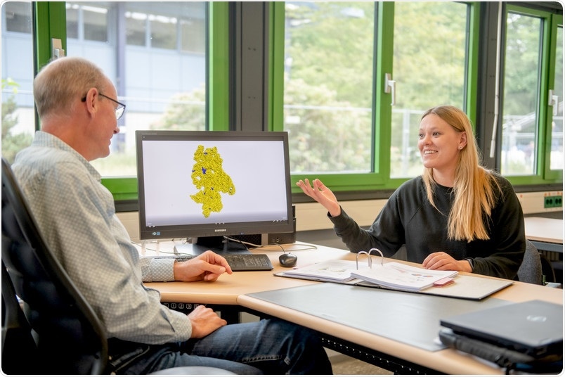
Franz Narberhaus and Vivian Brandenburg are discussing one of the deciphered RNA structures. Image Credit: Ruhr-Universität Bochum, Marquard.
Earlier, this had to be performed separately for every molecule. Apart from their precise composition, their structure is essential for the function of the RNAs. The researchers have explained the latest high-throughput structure mapping technique, called Lead-Seq that stands for lead sequencing, in the Nucleic Acids Research journal, published online on May 28th, 2020.
At RUB, Christian Twittenhoff, Vivian Brandenburg, Francesco Righetti, and Professor Franz Narberhaus from the Chair of Microbial Biology partnered with the bioinformatics team led by Professor Axel Mosig at RUB and the research team headed by Professor Petra Dersch at the University of Münster, formerly from the Helmholtz Centre for Infection Research in Braunschweig.
No structure—no function
Genetic data is stored in double-stranded DNA in all living cells, and this is transcribed into single-stranded RNA, which then acts as a blueprint for proteins. But RNA is a linear copy of the genetic information and also usually folds into complex structures. The combination of partly folded double-stranded and single-stranded regions is very crucial for the stability and function of RNAs.
If we want to learn something about RNAs, we must also understand their structure.”
Franz Narberhaus, Professor, Chair of Microbial Biology, Ruhr-Universität Bochum
Lead ions reveal single-stranded RNA positions
Through lead sequencing, the authors have introduced a technique that allows concurrent analysis of all RNA structures in a bacterial cell. In this process, the scientists leveraged the fact that lead ions break down the strands in single-stranded RNA segments; folded RNA structures, that is, double strands, still remain untouched by lead ions.
With lead sequencing, the scientists were able to divide the single-stranded RNA areas at haphazard locations into smaller fragments, subsequently transcribed them into DNA, and finally sequenced them. Thus, the beginning of every DNA sequence corresponded to a previous strand break in the RNA.
This tells us that the corresponding RNA regions were present as a single strand.”
Franz Narberhaus, Professor, Chair of Microbial Biology, Ruhr-Universität Bochum
Predicting the structure using bioinformatics
Later, Vivian Brandenburg and Axel Mosig employed bioinformatics to analyze the data on the single-stranded RNA parts achieved in the experiments.
We assumed that non-cut RNA regions were present as double strands and used prediction programs to calculate how the RNA molecules must be folded. This resulted in more reliable structures with the information from lead sequencing than without this information.”
Vivian Brandenburg, Researcher, Chair of Microbial Biology, Ruhr-Universität Bochum
This method allowed the scientists to determine the structures of scores of RNAs of the bacterium Yersinia pseudotuberculosis at the same time. The scientists then compared the outcomes obtained by lead sequencing of certain RNA structures with outcomes obtained through conventional techniques—they were both the same.
New RNA thermometers discovered
The scientists performed their experiments at 25 °C and 37 °C, because certain RNA structures change based on the temperature. Using the so-called RNA thermometers, bacteria like the diarrhea pathogen Yersinia pseudotuberculosis can find out whether they are inside the host. The team identified the previously known RNA thermometers and also discovered many new ones through lead sequencing.
It took five years to establish lead sequencing. “I’m happy to say that we are now able to map numerous RNA molecules in a bacterium simultaneously. One advantage of the method is that the small lead ions can easily enter living bacterial cells. We therefore assume that this method can be used universally and will in future facilitate the detailed structure-function analysis of bacterial RNAs,” Franz Narberhaus concluded.
Source:
Journal reference:
Twittenhoff, C., et al. (2020) Lead-seq: transcriptome-wide structure probing in vivo using lead (II) ions. Nucleic Acid Research. doi.org/10.1093/nar/gkaa404.