The centriole is the organelle of interest if individuals want to learn the fundamental mechanisms of cellular division and motility.

Image Credit: © 2020 EPFL/Hillary Sanctuary.
Every cell has two centrioles that help isolate chromosomes at the time of cell division. These unique organelles are multi-molecular machines made up of an unlimited number of proteins and contain an invisible code of post-translational modifications (PTMs) that account for their flexibility or rigidity, which, consequently, may help describe the function of centrioles.
The underlying structure of centrioles is identified based on earlier studies performed mostly through electron microscopy. Since PTMs are invisible under the electron microscope, it is not known how they look like.
EPFL biophysicists have now developed an improved super-resolution fluorescence microscope technology that helps one to have an in-depth picture of these nanoscale structures, both in situ and isolated.
As anticipated, the centrioles are molded similar to ridged bullets, that is, they are cylindrical with nine lengthwise ridges, with their diameter tapering off in one end.
Considering this high level of organization, the researchers were amazed to find that one PTM essentially twists around these lengthwise ridges. The study results were recently published in the Nature Methods journal.
The symmetries of multi-molecular machines often explain how they can perform diverse functions. PTMs can form a special code that tells proteins where to dock, but can also stabilize the centriole while forces are pulling during division.”
Suliana Manley, Biophysicist and Head, Laboratory of Experimental Biophysics, EPFL
Suliana Manley continued, “We still don’t know why the twist is there, but it offers a clue to how centrioles work. Our study underlines that super-resolution microscopy is an important partner to electron microscopy for structural biology.”
Improved super-resolution imaging techniques
Centrioles are around 100 times smaller than a mammalian cell and a 1000 times smaller than a human hair. Hence, observing the centrioles inside living cells called for enhanced super-resolution microscope technology that employs light to examine specimens, as the techniques tend to be very slow for structural studies.
Dora Mahecic, a Ph.D. student in the Laboratory of Experimental Biophysics, enhanced the illumination design to boost the size of images that could be captured by their microscope by supplying light more evenly over the field of view.
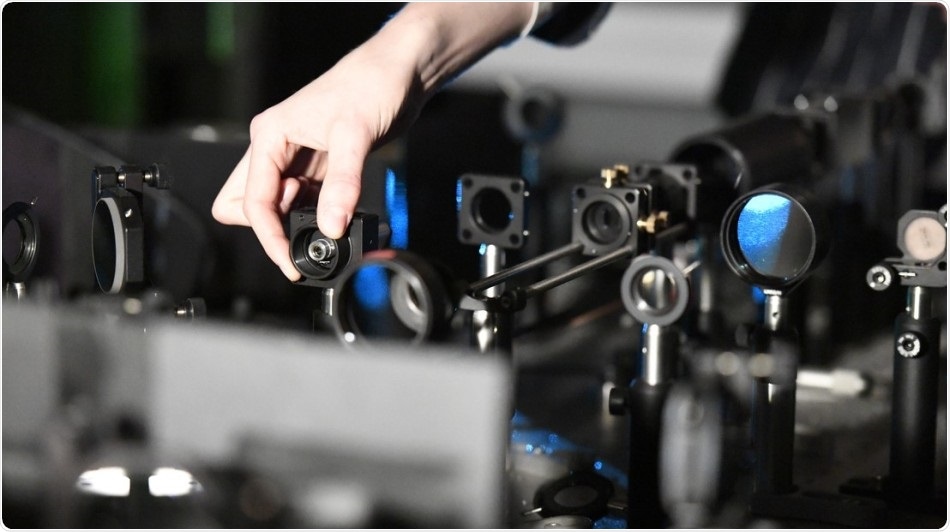
The microscope—a super-resolution fluorescence microscope—is completely different from the standard optical microscope that one would observe in an introductory biology class. It is, in fact, an intricate setup of carefully aligned lenses and mirrors that shape and supply laser light into the specimen.
The biophysicists integrated this setup with sophisticated sample preparation that employs physical magnification of the fluorophores and sample to form proteins—the building blocks of life—to re-emit light.
This unique super-resolution technology could be employed to various other structures inside the cell, such as mitochondria, or to examine other multi-molecular machines, like viruses.
Super-resolution microscopy reveals a twist inside of cells
Video Credit: EPFL.
Source:
Journal reference:
Mahecic, D., et al. (2020) Homogeneous multifocal excitation for high-throughput super-resolution imaging. Nature Methods. doi.org/10.1038/s41592-020-0859-z.