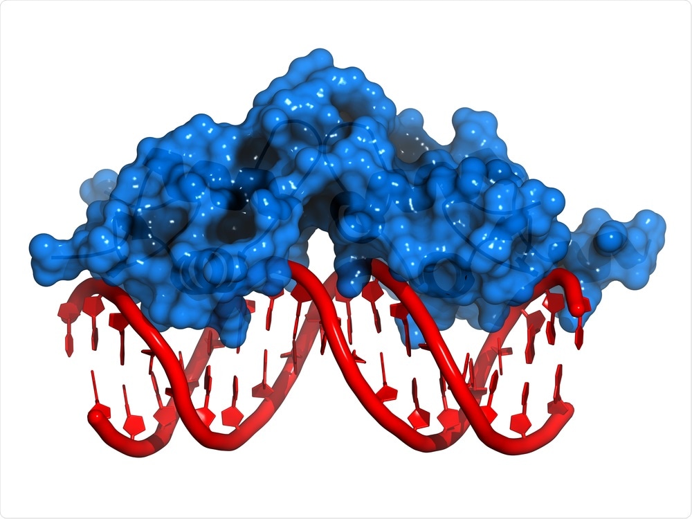
Life relies on a complex game of hide-and-seek that occurs in the interior of the cell. Now, a new study, recently published in the Nature journal, provides a new understanding of the mechanisms through which DNA-binding proteins look for the genome for their particular binding sites.

Image Credit: StudioMolekuul/Shutterstock.com
The double-helical molecule DNA stores the entire instructions that must be followed by a cell to maintain itself. The data is encoded inside the particular sequential order of genetic letters (that is, DNA base pair sequence). The correct implementation of the crucial instructions preserved in this genetic code is based on the potential of proteins to detect and selectively attach to particular DNA sequences.
These proteins comprise transcription factors, which play a major role by turning genes on and off by attaching to the particular transcription factor binding sites. If these DNA target sites are not engaged at the right time and the right place, it would have catastrophic consequences for cellular life—genes would no longer be turned on when required, while others may never switch off.
With regard to a transcription factor, identifying its specific binding site is more like finding the proverbial needle (that is the short stretch of DNA, usually around 12 genetic letters only) in a haystack (the genome, spanning from millions to billions of letters based on the organism).
While researchers have widely studied this so-called search problem, several proteins employ a process known as facilitated diffusion to speed up their search. In this case, a protein goes through three-dimensional (3D) diffusion, also called Brownian motion, until it haphazardly encounters a DNA molecule.
If the collision site does not match with the correct binding site, the protein can experience one-dimensional (1D) diffusion by haphazardly sliding to and fro along with the DNA prior to unbinding and returning to 3D diffusion.
Over the years, researchers have already established that the 1D sliding process speeds up the search, but the accurate mechanism of 1D sliding has continued to be elusive.
In the latest research, headed mutually by Uppsala University scientists Sebastian Deindl and Johan Elf, the 1D sliding mechanism plays the most important role.
The molecular mechanism of the scanning process has been poorly understood, and it has remained a great mystery how transcription factors manage to slide fast on non-specific DNA sequences, yet at the same time bind efficiently to specific targets.”
Emil Marklund, Joint First Author and PhD Student, Department of Cell and Molecular Biology, Uppsala University
To address these questions, both research groups created new fluorescence microscopy imaging techniques to visualize individual transcription factor proteins that slide along the DNA in real-time as they look for and adhere to the exact binding site.
It is exciting that we were able to develop new imaging approaches to directly observe, for the first time, if and how often the sliding protein fails to recognize and slides past its binding site.”
Sebastian Deindl, Researcher, Department of Cell and Molecular Biology, Uppsala University
It was found that the sliding protein is rather sloppy and often overlooks its target site. To gain a better understanding of how the sliding protein analyzes the surface of the DNA, a new method of monitoring and capturing extremely fast movies of the quickly sliding protein had to be created.
The protein rapidly searches the DNA—10 base pairs are scanned in about 100 µm—1 µm is equivalent to one-millionth of a second. The scientists realized that they have to perform relatively faster quantifications than anyone had done before to explore how the protein looks for the DNA surface on such length and timescales.
With new microscopy techniques, the researchers could trace the helical route of the sliding protein around the DNA molecule.
It’s great that we can push the dynamic observation of bimolecular interactions to the sub-millisecond time scale—this is where the chemistry of life happens.”
Johan Elf, Researcher, Department of Cell and Molecular Biology, Uppsala University
It turned out that the sliding protein does not rigorously track the path provided by the helical geometry of the DNA molecule itself, and instead slips out of its track quite often by making short hops.
“By hopping, the protein trades thorough scanning for speed, so it can scan DNA faster. This is a really smart choice by the protein since it will find the target twice as fast using this search mechanism,” Marklund concluded.
Source:
Journal reference:
Marklund, E., et al. (2020) DNA surface exploration and operator bypassing during target search. Nature. doi.org/10.1038/s41586-020-2413-7.