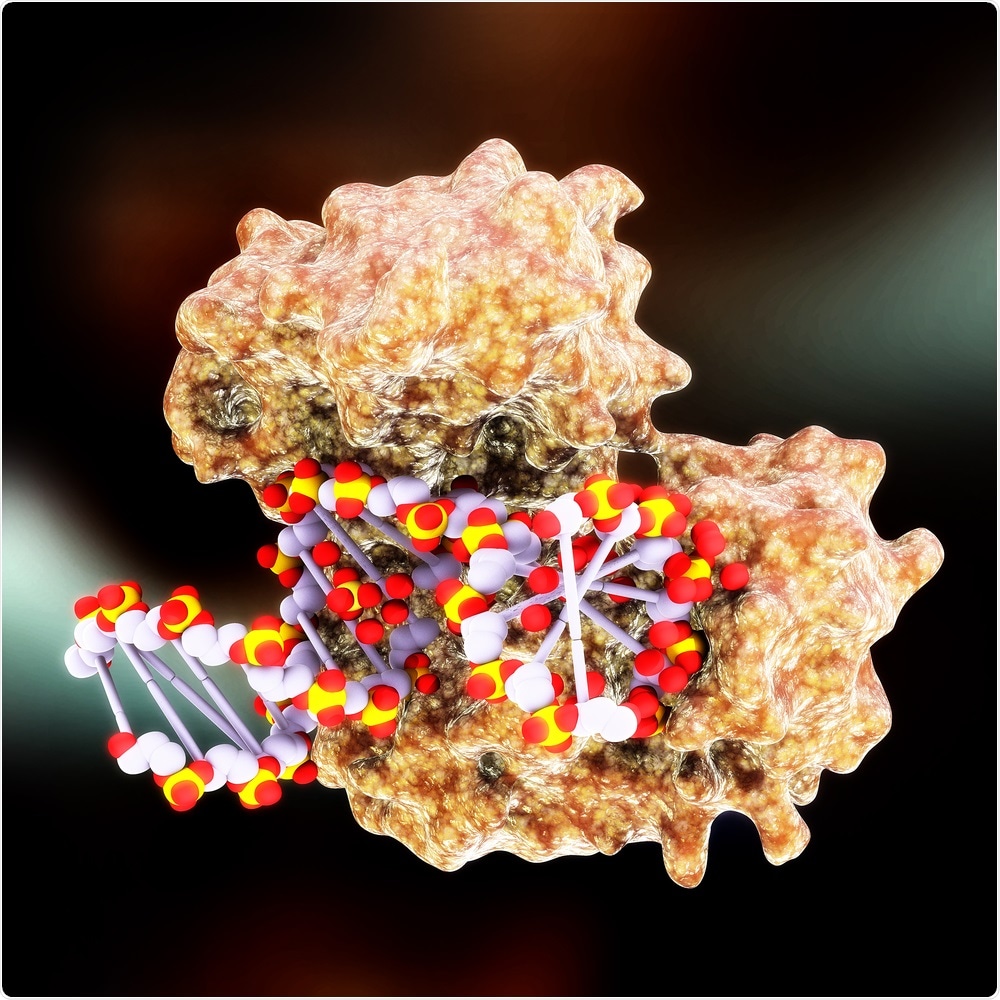
In a first-of-its-kind study, a team of researchers from The Mount Sinai Hospital has revealed the mechanism and 3D structure of a complex enzyme that guards cells against persistent DNA damage. This latest discovery creates opportunities to identify novel therapeutics for treating chemotherapy-resistant cancers.

Image Credit: MichaelTaylor3d/Shutterstock.com
In this study, the team used sophisticated cryo-electron microscopy technique to gain a deeper understanding of the enzyme called DNA polymerase ζ (Pol ζ), whose mechanism and architecture continued to be a mystery to investigators for many years.
The study was published in the Nature Structural & Molecular Biology journal in August 2020.
Resolving the structure of the complete Pol ζ enzyme at near-atomic resolution allows us to address long-standing questions of how this unique polymerase replicates through daily DNA-damaging events, while also providing a template for designing drugs against cancers that are refractory to conventional chemotherapeutics.”
Aneel Aggarwal, PhD, Study Lead Author and Professor, Department of Pharmacological Sciences, Icahn School of Medicine at Mount Sinai
The crucial enzyme Pol ζ enables cells to fight over 100,000 events that impair the DNA. Such events occur every day, right from regular metabolic activities to environmental intrusions, such as industrial carcinogens, ionizing radiation, and ultraviolet light.
At The Mount Sinai Hospital, the research team, including Radhika Malik, PhD, Assistant Professor of Pharmacological Sciences and the study’s first author, learned how the cells are protected by the Pol ζ enzyme from both man-made and natural environmental and cellular stresses through a complex structure of four different kinds of proteins that link to one another in a daisy chain-like, or pentameric, configuration.
It is believed that this architecture will offer useful insights to investigators for the upcoming development of medications designed to block the Pol ζ enzyme when treating certain types of cancers, such as ovarian, prostate, and non-small-cell lung cancers, which often become impervious to chemotherapy when it is used early in patients.
The reason behind this resistance is that chemotherapies, similar to cisplatin, actually rely on their DNA-damaging effects. Therefore, inhibiting and blocking the role of the Pol ζ enzyme renders the tumor cells more susceptible to the therapeutic effect of chemotherapy.
The development of effective inhibitors has been hampered in the past by a lack of structural information on Pol ζ. Our work now offers a much clearer picture, and we expect these new insights will spur efforts by scientists around the world to create effective new therapies. For the thousands of patients with tumors that are resistant to chemotherapy, these findings could prove to be particularly valuable by meeting an unfulfilled need in their battle against cancer.”
Aneel Aggarwal, PhD, Study Lead Author and Professor, Department of Pharmacological Sciences, Icahn School of Medicine at Mount Sinai
There was no progress over the years and this was largely because structural analyses of the Pol ζ enzyme were restricted by the unattainability and low yields of well-diffracting crystals.
Dr Aggarwal and his research team resolved that issue by using advanced cryo-electron microscopy. This technique, which helps image quickly frozen molecules in solutions, is redefining the whole field of structural biology via its high-resolution images of complex molecules.
Source:
Journal reference:
Malik, R., et al. (2020) Structure and mechanism of B-family DNA polymerase ζ specialized for translesion DNA synthesis. Nature Structural & Molecular Biology. doi.org/10.1038/s41594-020-0476-7.