Researchers are still clueless about how the neural networks are linked together to make functional circuits. For a long time, this has been a prevailing question in the field of neuroscience.

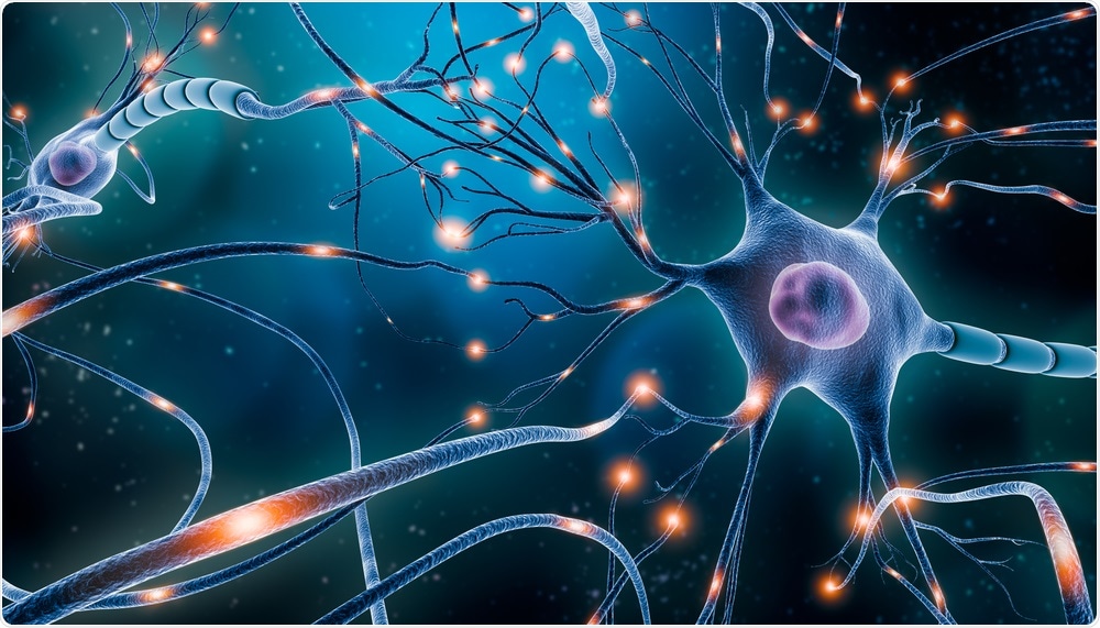
Neurons. Image Credit: MattLphotography/Shutterstock.com
To answer this underlying question, scientists from Boston Children’s Hospital and Harvard Medical School have designed a new method to analyze these circuits and, in the process, get to know more about the relationships between them.
Neural networks are extensive, but the connections between them are really small. So, we have had to develop techniques to see them in extremely high-resolution over really large areas and volumes.”
Wei-Chung Allen Lee, PhD, F.M. Kirby Neurobiology Center, Boston Children’s Hospital and Harvard Medical School
To achieve this, Lee’s team came up with a better process for large-scale electron microscopy (EM)—a method that was initially created in the 1950s with the help of accelerated beams of electrons to view tiny structures.
But the problem with electron microscopy is that because it provides such high image resolution, it has been difficult to study whole neural circuits. To improve the technique, we developed an automated system to image at high-resolution, but at the scale to encompass neuronal circuits.”
Wei-Chung Allen Lee, PhD, F.M. Kirby Neurobiology Center, Boston Children’s Hospital and Harvard Medical School
Lee’s laboratory is interested in understanding how neural circuits underlie behavior and function. An article explaining the new findings was published in the Cell journal.
GridTape: Automated, Faster, Cheaper Electron Microscopy Technique
In a conventional EM technique, scores of tissue samples are manually collected onto a grid. The tissue is cut into sections, which measure 40-nm thick, that is, around 1000 times thinner than a single strand of human hair.
The method automates the process of sample collection, assigning a barcode to all the sections, and placing them on a conveyor belt that could subsequently be fed via an electron microscope, for example, a movie projector. One major benefit of this method is that all neurons are labeled in every tissue section.
As the electrons pass through each section, we can image each neuron in fine detail. And because all of the sections are labeled with a barcode we know exactly where each of these sections comes from so we can reconstruct the circuits.”
Wei-Chung Allen Lee, PhD, F.M. Kirby Neurobiology Center, Boston Children’s Hospital and Harvard Medical School
“This new technique allows us to do electron microscopy faster and in an automated way, with high quality, yet at a reasonable price,” added Lee.
In their article, the researchers have provided GridTape instrumentation software and designs to make large-scale EM affordable and accessible to the larger scientific research community.
Fruit fly spinal cord: a case study
The researchers applied their GridTape technique to investigate the ventral nerve cord of the Drosophila melanogaster fruit fly, which is almost the same as the spinal cord. The ventral nerve cord contains all the circuits used by the fly to move its limbs. As such, the team is aiming to create a detailed map of the neuronal circuits that regulate motor function.
“By applying this method to the entire nerve cord, we were able to reconstruct all of its motor neurons, as well as a large population of sensory neurons,” Lee added.
During the process, the researchers identified a certain kind of sensory neuron in the fly, believed to detect changes in load, such as body weight.
“These neurons are very large, relatively rare in number, and they make direct connections onto motor neurons of the same type on both sides of the body. We believe this may be a circuit that helps stabilize body position,” Lee further added.
Based on the new study, the researchers generated a map of over 1,000 sensory and motor neuron reconstructions that are available on an open registry.
“It allows anyone in the world to access this data set and look at any neuron that they're interested in and ask who they’re connected to,” added Lee.
Future applications
Now, with the potential to plot an increasing number of larger neural circuits, Lee hopes that the new method may prove invaluable for analyzing neuronal circuits in bigger brains and for testing predictions about the behavior and function of neurons.
Lee’s group is presently investigating the method in mice, while other investigators in Japan and the United Kingdom are using the method across numerous animal systems.
Besides, the technology also has more expensive potential applications, where huge numbers of samples had to be imaged at an extremely high resolution.
“So, in principle, various forms of electron microscopy could be advanced by using this technique if people need to generate lots and lots of data,” added Lee, such as DNA sequencing using EM, or cryogenic EM, to resolve the structures of proteins.
Source:
Journal reference:
Phelps, J. S., et al. (2021) Reconstruction of motor control circuits in adult Drosophila using automated transmission electron microscopy. Cell. doi.org/10.1016/j.cell.2020.12.013.