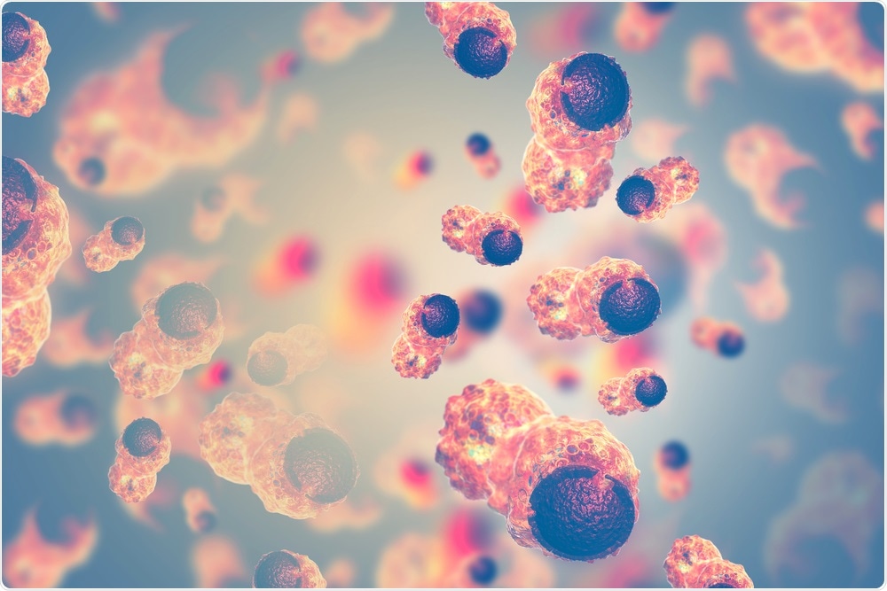
A lot of biological mechanisms go wrong in cancer—for example, molecular processes change significantly, genes mutate, and cells proliferate in an uncontrollable manner to form completely new tissues that are referred to as tumors.

Cancer Cells. Image Credit: crystal light/Shutterstock.com
In fact, many things go awry at different levels, and this complication partly makes it very hard to study and treat cancer. Hence, it is no surprise why cancer researchers pay more attention to the genome, where all types of cancers start. If scientists can understand the mechanisms that occur at the DNA level, then it might become possible to treat and even prevent cancers completely in the days to come.
This determination has spurred a research team from EPFL and the University of Lausanne (UNIL) to make a groundbreaking discovery relating to a crucial genetic abnormality that takes place in cancer.
Working closely together, the teams of Elisa Oricchio from EPFL and Giovanni Ciriello from UNIL have applied an innovative algorithm-based technique to investigate how tumor cells re-assemble the 3D structure of their DNA to boost the activity of cancer-promoting genes, known as “oncogenes.” The study has been published in two journals, Nature Communications and Nature Genetics.
The study targets chromosomes, in which the human DNA is packaged, and the way the chromosomes are arranged in the constricted space of the cell nucleus. Considering that every single one of the infinite number of cells in the human body contains around 2 m of DNA, it can be understood that humans evolved mechanisms to store it correctly.
In these mechanisms, DNA is wound around histones, which are specialized proteins similar to a string spooling around a yoyo.
The resulting well-protected and super-packed DNA-protein complex is known as chromatin. Numerous chromatin units constitute the structures referred to as chromosomes. Generally, every cell carries 23 chromosomes and a couple of copies for every chromosome; however, the organization and structure of these chromosomes change in the cancer cells.
For instance, a part of a copy of chromosome 8 can be linked to a copy of chromosome 14. Furthermore, a chromosome can assume a more compact or relaxed structure, which relies on chemical changes known as “epigenetic marks.”
The team investigated how the variations in certain epigenetic marks alter the structures of chromosomes and the expression of genes that support the growth of tumors or oncogenes.
Giovanni Ciriello’s research group from UNIL designed an innovative algorithmic method, known as Calder (named after the American sculptor Alexander Calder) to monitor how genomic areas are placed with regards to one another in the nucleus.
We used Calder to compare the spatial organization of the genome in more than a hundred samples. But this organization is not static and, just like Alexander Calder's mobile sculptures, it can rearrange its pieces.”
Giovanni Ciriello, University of Lausanne
The team used the Calder technique to track chromatin regions that “moved” from one region of the nucleus to another because of varying epigenetic marks. In the meantime, Oricchio’s research team from EPFL used the same Calder technique to track the variations of the chromatin 3D structure in both B-cell lymphoma and normal cells.
The researchers observed that particular epigenetic changes in the lymphoma cells cause the regions of chromatin to be repositioned in different nucleus areas, which resulted in new local interactions that overstimulate the expression of oncogenes.
The researchers also discovered that when a pair of fragments from varied chromosomes are spliced and swapped, they take on a 3D structure that can be distinguished from the normal copies. Most significantly, such changes of 3D structure correlate with different epigenetic marks and trigger high expression of genes that promote the expansion of tumor cells.
Most of the time we think of our DNA as a long, linear molecule, and it's only recently that we started to understand how its 3D organization is altered in cancer cells. Considering the spatial organization of DNA in the nucleus provides a new lens to understand how tumor cells originate, and how therapeutic modulation of epigenetic marks can block tumor progression.”
Elisa Oricchio, EPFL
Source:
Journal reference:
Sungalee, S., et al. (2021) Histone acetylation dynamics modulates chromatin conformation and allele-specific interactions at oncogenic loci. Nature Genetics. doi.org/10.1038/s41588-021-00842-x.