Viruses infect cells, causing changes to the cell nucleus. These changes can be visualized through fluorescence microscopy. Researchers from the University of Zurich have now used fluorescence images from live cells to train an artificial neural network to reliably detect cells that are infected by herpes viruses or adenoviruses. Through this procedure, severe acute infections can also be identified at an early stage.

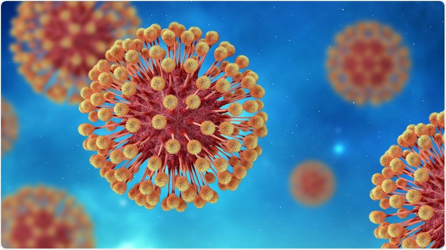
Herpes viruses can multiply in skin and nerve cells for years without causing disease symptoms, but can also cause sudden, violent infections. Image Credit: iStock.com/photoman
Adenoviruses can infect the respiratory tract in humans, while herpes viruses can infect both the skin and the nervous system. In a majority of cases, the immune system suppresses the viruses and hence does not lead to the production of new viral particles. But both herpes viruses and adenovirus can cause persistent infections that cannot be fully suppressed by the immune system. This leads to the production of viral particles for many years.
The same viruses can even lead to spontaneous, violent infections where the affected cells produce large amounts of viruses and cause the infection to spread quickly. This can lead to serious acute diseases of the nervous system or the lungs.
Automatic detection of virus-infected cells
For the first time, a research team led by Urs Greber, Professor from the Department of Molecular Life Sciences at the University of Zurich (UZH), has now revealed that a machine-learning algorithm can detect the cells infected with adenoviruses or herpes, mainly based on the fluorescence of the cell nucleus.
Our method not only reliably identifies virus-infected cells but also accurately detects virulent infections in advance.”
Urs Greber, Professor, Department of Molecular Life Sciences, University of Zurich
The team believes that this latest development has many different applications, like predicting how human cells respond to other microbes or viruses. Greber added, “The method opens up new ways to better understand infections and to discover new active agents against pathogens such as viruses or bacteria.”
The new technique combines fluorescence microscopy in living cells with deep-learning processes. The adenovirus and herpes virus formed within an infected cell alter the organization of the nucleus and such changes can be seen under a microscope.
The researchers designed a deep-learning algorithm—a kind of artificial neural network—that automatically spots these changes. They trained the network with a huge set of microscopy images through which it learns to detect patterns that are typical of infected or uninfected cells.
After training and validation are complete, the neural network automatically detects virus-infected cells.”
Urs Greber, Professor, Department of Molecular Life Sciences, University of Zurich
Reliably predicting severe acute infections
The researchers also demonstrated that the deep-learning algorithm can identify both acute and severe infections with an accuracy of 95% and up to 24 hours in advance. Pictures of living cells from lytic infections, where the viral particles multiply quickly and the cells dissolve, as well as pictures of persistent infections, where viruses are produced constantly but only in limited quantities, served as training material.
While the method is highly precise, it is still not clear which traits of the infected cell nuclei are detected by the artificial neural network to differentiate the two phases of infection. But even without this information, the team was able to investigate the biology of infected cells in more detail.
The researchers have already noticed some differences: The nucleus’s internal pressure is greater during virulent infections than during persistent phases of infections. In addition, the viral proteins accumulate more quickly in the nucleus in a cell with lytic infection.
We suspect that distinct cellular processes determine whether or not a cell disintegrates after it is infected. We can now investigate these and other questions.”
Urs Greber, Professor, Department of Molecular Life Sciences, University of Zurich
Source:
Journal reference:
Andriasyan, V., et al. (2021) Microscopy deep learning predicts virus infections and reveals mechanics of lytic infected cells. iScience. doi.org/10.1016/j.isci.2021.102543.