Researchers have achieved several discoveries in the past, thanks to the availability of better, more accurate measurement methods, which help obtain data from phenomena unexplored earlier.

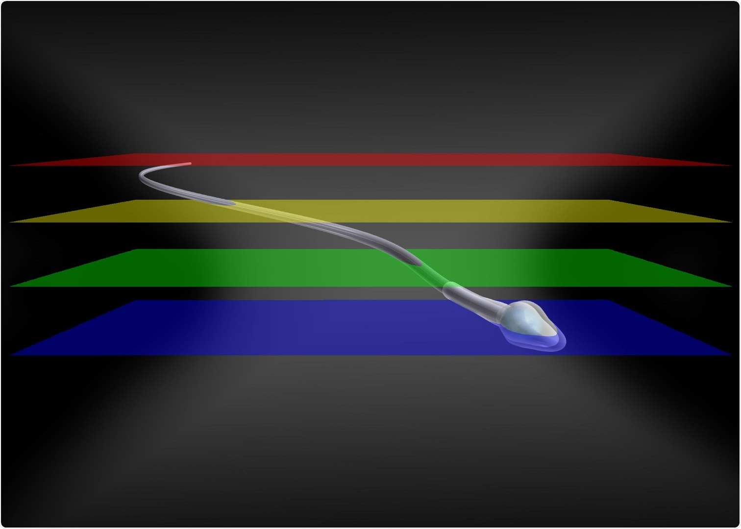
A new method provides novel insight into the dynamics of flagellar beating and its connection to the swimming behavior of sperm. Image Credit: © René Pascal.
For instance, high-resolution microscopy has started changing the perspectives of cell function and dynamics drastically. Scientists from the ImmunoSensation2 Cluster of Excellence at the University of Bonn, the University Hospital, and the research center caesar have come up with a method that enables the use of multi-focal images to reconstruct the movement of fast biological processes in 3D.
The study was published recently in the Nature Communications journal.
Numerous biological processes occur within a few milliseconds on a nano- to millimeter scale. Although standard techniques like confocal microscopy are well suited for accurate 3D recordings, they lack the temporal or spatial resolution to resolve fast 3D processes and need labeled samples.
Image acquisition at high frame rates is crucial for various investigations in biology to record and comprehend the principles that regulate cellular functions or fast animal behaviors. The difficulty faced by researchers is comparable to following a thrilling tennis match: At times, it is not feasible to follow the fast-moving ball with precision, or the ball is not found before it is already out of bounds.
Previous techniques were not useful for researchers to track the shot as the image was blurred or the object of interest was just no longer in the field of view once the picture was captured. Standard multifocal imaging techniques enable high-speed 3D imaging but are restricted by the compromise between a large field of view and high resolution. Moreover, they often need bright fluorescent labels.
The newly developed technique enables, for the first time, multifocal imaging with both a high spatio-temporal resolution and a large field of view. As part of this study, the researchers quickly and precisely tracked the movement of non-labeled spherical and filamentous structures.
As illustrated in the new study, the new technique remarkably offers new insights into the dynamics of flagellar beating and its relation to the swimming behavior of sperm. This link has been viable since the researchers could precisely record the flagellar beat of free-swimming sperm in 3D over a longer time and concurrently follow sperm trajectories of individual sperm.
Besides, the researchers unraveled the 3D fluid flow around the beating sperm. These insights not just pave the way to comprehend the causes of infertility but could even be used in what is called “bionics,” or the transfer of principles that occur in nature to technical applications.
Scientists at the ImmunoSensation2 Cluster of Excellence are already able to employ the new method—not just to visualize sperm. This technique could even be employed to establish the 3D flow maps resulting from the beating of motile cilia.
The beats of motile cilia are similar to those of the sperm tail and transport fluid. Cilia-induced flow has a vital role in the ventricle of the brain or in the airways where it acts to carry mucus out of the lungs and into the throat—this is how pathogens are carried out and warded off.
The multi-focal imaging method discussed in this study is economical, can be easily executed, and does not depend on object labeling. The scientists claim that the new technique could be used even in other fields, and they envision various other prospective applications.
Source:
Journal reference:
Hansen, J. N., et al. (2021) Multifocal imaging for precise, label-free tracking of fast biological processes in 3D. Nature Communications. doi.org/10.1038/s41467-021-24768-4.