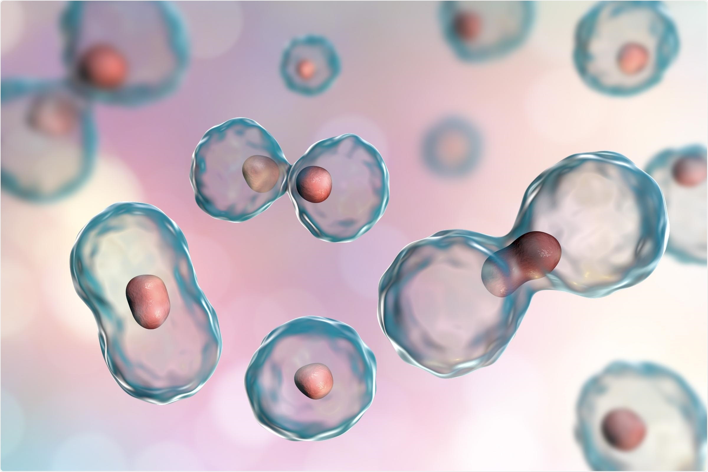
The hereditary material, DNA, interacts with proteins present in the cell nuclei and forms a tightly packed complex with proteins called “chromatin”. The molecular architecture of the chromatin controls access to genes, and thus ascertains the genes to be activated in various cell types.

Image Credit: © Kateryna_Kon—stock.adobe.com.
Numerous chemical modifications of proteins known as histones—performing a decisive role in the structuring of chromatin—function as markers that can either inhibit or activate gene expression. However, it is not yet completely understood how these epigenetic modifications are regulated during development.
Recently, scientists headed by LMU molecular biologist Ralph Rupp demonstrated that the mechanism of cell division itself has an effect on which modifications become dominant. In quickly dividing cells, levels of inhibitory modifications are decreased, thus raising the probability of silenced genes being reactivated.
This phenomenon takes place in embryonic cells, and mostly in adult stem cells and precursor cells. The observations were published in the PLOS Biology journal.
Histone modification codes for vital information that helps assure the orderly progression of early development. Rupp’s team used an established experimental model—the tadpole stage of the clawed toad Xenopus laevis—to demonstrate the various histone modifications become dominant in different stages of embryonic development.
To our great surprise, we found that the cell cycle—the sequence of events that takes place in cells in the interval between one division and the next—has an impact on the profile of modifications present in the chromatin. When cells switch between a quiescent, resting state and a proliferative state—in which cells divide more often—certain epigenetic markers are selectively altered.”
Ralph Rupp, Molecular Biologist, Ludwig-Maximilians-Universität München
The rate of cell proliferation regulates repressive histone modifications
The fact that the rate of cell proliferation controls repressive histone modifications holds particularly for repressive modifications, which silence genes.
Such modifications are apparently very sensitive to what is known as the S-phase dilution effect.”
Ralph Rupp, Molecular Biologist, Ludwig-Maximilians-Universität München
This effect arises from the fact that during cell division, chromatin along with the DNA should be replicated. The prevalent histone proteins get equally distributed between the replicated DNA strands, while the newly produced histones—which are not yet modified—fill in the gaps.
Thus, the degree of histone modification of the daughter chromosomes instantly after cell division is only half as big as it was before DNA replication.
The findings on the clawed toad revealed that the rate of cell proliferation exceeds the rate at which repressive histone modifications are produced in chromatin. A major indication is that these are the genes that were already inactivated and return to a less repressed state when a cell determines to divide again. This shows that the properties of any cell are more pliable than anticipated.
In contrast, in resting cells the total number of repressive markers increases – and at some stage it reaches a level at which specific genes become permanently inactivated.”
Ralph Rupp, Molecular Biologist, Ludwig-Maximilians-Universität München
The researchers state that similar to embryonic cells, this process also performs a vital role in adult stem cells and precursor cells. They further imply that it can be employed as a means of cell reprogramming to make them more effective for specific therapeutic purposes.
Source:
Journal reference:
Pokrovsky, D., et al. (2021) A systemic cell cycle block impacts stage-specific histone modification profiles during Xenopus embryogenesis. PLoS Biology. doi.org/10.1371/journal.pbio.3001377.