G protein-coupled receptors (GPCRs), membrane proteins that are the target of one-third of approved drugs, are being studied by scientists at St. Jude Children’s Research Hospital and Columbia University/New York State Psychiatric Institute.
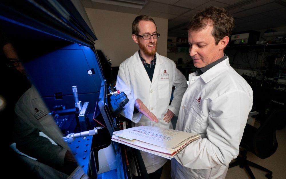
 Dr Daniel Terry and Dr Scott Blanchard, co-authors of a new publication in Cell at St. Jude Children’s Research Hospital. Image Credit: St. Jude Children’s Research Hospital.
Researchers used single-molecule imaging methods to obtain a new understanding of the way by which GPCRs convey cellular signals. By changing the way drugs stimulate certain pathways, the research might assist in the creation of new drugs. The study was published in the journal Cell.
Around 800 GPCRs are encoded in the human genome, and they are expressed throughout the body and control a variety of physiological processes. GPCRs are key therapeutic targets for a lot of diseases.
When agonists activate GPCRs, they send signals inside the cell (which are chemical signals such as neurotransmitters, cytokines, hormones, or synthetic drugs). β-arrestins are proteins that stick to GPCRs to inhibit G protein-mediated signaling while simultaneously triggering a number of downstream signaling pathways.
A particular region of the GPCR, generally its tail, is involved in the interaction of a GPCR with β-arrestin. The phosphorylation of this tail is activated-dependent. The GPCR tail is phosphorylated and binds into a groove on β-arrestin. This groove is filled by β-arrestin’s own C-terminal tail when it is not associated with a GPCR. The researchers sought to know how the C-terminal tail of the β-arrestin is released to create room for the phosphorylated GPCR tail.
We are trying to understand the process by which information is transmitted from the outside to the inside of the cell through GPCRs and, specifically, whether this information transfer occurs by way of detectable conformational [shape] changes in GPCRs and their binding partners. Single-molecule imaging is a way to directly measure molecular-scale conformational changes that is very insightful and often more easily interpreted than other approaches.”
Scott Blanchard, PhD, Study Co-Corresponding Author, Department of Structural Biology, St. Jude Children’s Research Hospital
A tale of two tails
The conformational dynamics (shape variations) of the β-arrestin C-terminal tail were studied using single-molecule fluorescence resonance energy transfer (smFRET). The majority of research in this field has used ensemble techniques, which average hundreds of thousands of proteins.
These averages do not reveal what a given protein is up to. The single-molecule technique, on the other hand, can give a direct proof of a protein’s activity and allow researchers to investigate how it evolves over time.
The findings reveal that the resting β-arrestin is generally autoinhibited in a stable condition. The C-terminal tail is securely linked to the groove in this form. An agonist must attach to the GPCR, which then “tickles” the β-arrestin to initiate the release of the C-terminal tail and create room for the phosphorylated GPCR tail.
The researchers were able to separate the contributions of the GPCR’s phosphorylated state from the agonist-activated state using smFRET, which is something that cannot be addressed in the cell. The capacity to distinguish between these states led to the revelation that the receptor tail has an autoinhibitory function that must be eased by agonist binding.
The strength and duration of GPCR signaling are controlled by the balance between autoinhibited and activated states, which influences physiological responses. The discoveries might help with drug development since they allow for a systematic examination of the effects of GPCR phosphorylation patterns and extents.
Now that we know receptors can both activate G-proteins and mediate signaling through β-arrestin, the hope is that the field can develop more specific pharmacotherapies by finding small molecules that preferentially activate one pathway or the other.”
Jonathan Javitch, MD, PhD, Study Co-Corresponding Author, Columbia University and the New York State Psychiatric Institute
Source:
Journal reference:
Asher, W. B., et al. (2022) GPCR-mediated β-arrestin activation deconvoluted with single-molecule precision. Cell. doi.org/10.1016/j.cell.2022.03.042.