Proteins are among the most significant and diverse biomolecules found in living organisms. These strings of amino acids, which take on complex 3-dimensional structures, are required for tissue growth and maintenance, the activation of thousands of biochemical events, and the immune system’s defense against invaders. They are the main targets for pharmaceutical medicines and play an important role in health and disease.
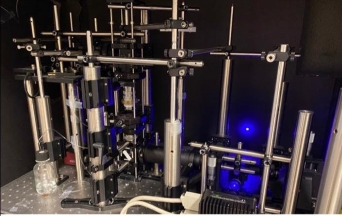
 The photograph shows the experimental setup for ESM microscopy. Image Credit: Arizona State University
Scientists have built complex technologies to examine and study proteins through enhanced microscopy, improved light detection, imaging software, and the incorporation of complex hardware systems to completely comprehend proteins and their various activities.
Shaopeng Wang and his colleagues at Arizona State University report a novel technology that offers to transform the imaging of proteins and other essential biomolecules, enabling these tiny structures to be viewed with exceptional clarity and using simpler ways than previous methods.
The method we report in this study uses normal cover glass instead of gold-coated cover glass, which has two advantages over our previously reported label-free single-protein imaging method. It is compatible with fluorescence imaging for in-situ cross-validation, and it reduces the light-induced heating effect that could harm the biological samples. Pengfei Zhang, an outstanding postdoctoral researcher in my group, is the technical lead of this project.”
Shaopeng Wang, Arizona State University
Wang is a member of the Biodesign Center for Bioelectronics and Biosensors as well as the School of Biological and Health Systems Engineering’s staff. The results of the group’s research are published in the recent edition of Nature Communications.
The new technology, called evanescent scattering microscopy (ESM), is built on an optical phenomenon known as total internal reflection, which was first discovered in antiquity. When the light goes through a high-refractive medium (such as glass) and into a low-refractive medium (like water), this happens.
When incident light is shifted away from perpendicular (relative to the surface), it finally finds the “critical angle,” causing all incident light to be reflected instead of flowing through the second medium. (Laser light is utilized to adequately expose biological samples.)
When cells or molecules like proteins adhere to a cover glass, an evanescent field is created, which can stimulate cells or molecules like proteins at the glass-water contact, allowing scientists to see them in incredible detail.
To better view biomolecules of concern, previous approaches often labeled them with fluorescent tags termed fluorophores. This method can obstruct the tiny interactions being detected and necessitates time-consuming sample preparation. The ESM method is a label-free imaging technique that does not require the use of fluorescent dye or gold coating on experiment slides.
Instead, the approach uses minor flaws in the cover glass’s surface to create images with razor-sharp contrast. The intrusion of evanescent light scattered by single-molecule samples and the rough surface of the cover glass is imaged to achieve this.
Evanescent wave scattering permits samples, such as proteins, to be examined at incredibly shallow depths, often less than 100 microns. ESM may now construct an optical slice with dimensions similar to a thin electron microscopy segment.
ESM was used to identify four model proteins in the new research: bovine serum albumin (BSA), mouse immunoglobulin G (IgG), human immunoglobulin A (IgA), and human immunoglobulin M (IgM).
In a series of tests, researchers noted protein-protein interactions, particularly fast binding and detachment of specific proteins. Studying binding kinetics is critical for developing medications that are both safer and more efficient. The scientists also utilized ESM to monitor structural changes in DNA, proving the novel method’s potency and applicability.
Source:
Journal reference:
Zhang, P., et al. (2022) Evanescent scattering imaging of single protein binding kinetics and DNA conformation changes. Nature Communications. doi.org/10.1038/s41467-022-30046-8.