Scientists at the Universities of Bonn and Osnabrück have identified a protein that when defective in flies results in motor dysfunction. Prior research has also shown the presence of the protein in Parkinson’s disease patients in people. However, it was unclear until recently what purpose it served in the cell. This question is now answered by the research. Science Advances has recently published the research, which also included the University Hospital Aachen.
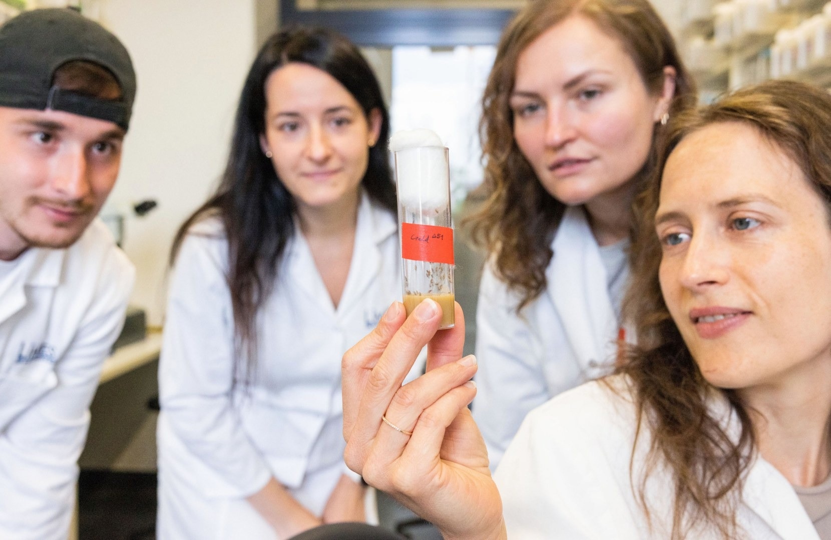
 A research team from the University of Bonn has studied the function of a protein in flies. From left to right: Santiago Maya, Dr. Gabriela Edwards Faret, Nicole Kucharowski, and study leader Dr. Margret Helene Bülow. Image Credit: Meike Böschemeyer/ University of Bonn.
The research teams examined a protein known as Creld for their investigation. Creld plays a significant role in the mammalian heart’s development, according to research from Bonn that was just reported.
We wanted to find out exactly what the protein does.”
Dr Margret Helene Bülow, Lecturer, LIMES Institute, University of Bonn
The scientists used genetically altered fruit flies from the genus Drosophila that are unable to develop Creld for their studies on these issues. The animals’ heart rates decreased in a certain manner, which is an indication of limited energy. They showed serious motor abnormalities as well. Energy is produced by the mitochondria, which serve as the cell’s power plants.
The nerve cells that give people their ability to move may die as a consequence of their dysfunction. Parkinson’s disease is the clinical diagnosis.
So Creld may play an important role not only in impaired heart function, but also in Parkinson’s disease. The findings of a recent analysis are consistent with this. It suggests that Creld production is often reduced in Parkinson’s patients.”
Dr Nicole Kucharowski, LIMES Institute, University of Bonn
Dr Kucharowski also worked with Marie Paradis on a significant portion of the study’s experiments.
But it remained unclear how exactly Creld and Parkinson’s may be related: The protein is not at all present in mitochondria. It can only be found in the endoplasmic reticulum (ER), a highly branching network of tubes that is responsible for producing a variety of chemicals in cells. From there, how may it affect the operation of cellular power plants?
Pesticide suspected of causing Parkinson’s disease
The researchers gave healthy fruit flies modest doses of a pesticide to find out (i.e., those that can form Creld). It has rotenone as an active component, which is thought to cause Parkinson’s disease in people.
Rotenone directly affects the mitochondria by obstructing an important stage in the synthesis of energy. After receiving the pesticide, the flies had motor abnormalities like those of Creld mutants. “We also found that their mitochondria are very often in contact with the ER,” Bülow says.
The researchers were able to demonstrate via further studies that certain classes of lipids are transferred from the ER to the mitochondria during this interaction. These so-called phospholipids restart the process by which rotenone inhibits the synthesis of energy. The mitochondria attempt to increase the energy supply again in this manner with help from the ER.
And the availability of Creld seems to be crucial for this transfer of phospholipids. In flies that cannot form Creld, phospholipids accumulate in the contact sites between ER and mitochondria. So they don’t get transported to the mitochondria, but accumulate.”
Dr Margret Helene Bülow, Lecturer, LIMES Institute, University of Bonn
Creld increases energy production in the cell
Creld is crucial for boosting the cell’s ability to produce energy. This is consistent with the finding that Drosophila mutants without Creld hardly create any hydrogen peroxide in their mitochondria—a waste product generated during the operation of the power plants. Hydrogen peroxide is a molecule.
Cells may be harmed by hydrogen peroxide. Up until this point, it was assumed that Parkinson’s patients created it excessively or that it was not properly disposed of. The motor function-related nerve cells would eventually get poisoned by this.
It is also feasible that another impact, such as the chronic energy shortage brought on by Creld destruction or underproduction, would lead to their extinction. Bülow, a member of the University of Bonn’s Transdisciplinary Research Area “Life and Health,” adds, “This is a thesis that we now need to investigate further.”
A successful partnership also contributed to the present success. As an example, crucial components of the study were completed at the University of Osnabrück. The study’s initial proponent, Dr Julia Sellin, recently relocated to the University Hospital in Aachen.
“The collaboration with Prof. Dr Christoph Thiele from the Cluster of Excellence Immunosensation2 here at the University of Bonn also went extremely well,” concludes Bülow.
Source:
Journal reference:
Paradis, M., et al. (2022) The ER protein Creld regulates ER-mitochondria contact dynamics and respiratory complex 1 activity. Science Advances. doi.org/10.1126/sciadv.abo0155.