Some cells travel quicker in thicker fluids, such as honey against water or mucus versus blood, because their ruffled edges detect the viscosity of their surroundings and adjust to boost their speed.
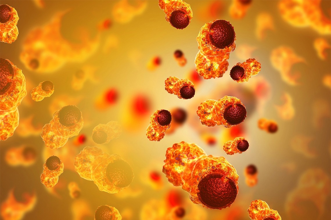
 A study co-authored by U of T researchers suggests some cells travel faster in thicker fluid by using “membrane ruffling to sense changes in extracellular fluid viscosity and to trigger adaptive responses.” Image Credit: Mohammed Haneefa Nizamudeen/iStock.
This was one of the surprising discoveries of a recent study published in Nature Physics by researchers at the University of Toronto, Johns Hopkins University, and Vanderbilt University.
The researchers’ findings in cancer and fibroblast cells—the type that frequently forms scars in tissues—suggest that the viscosity of a cell’s surrounding environment is a significant component of disease and may help explain tumor progression, scarring in cystic fibrosis-affected mucus-filled lungs, and the wound-healing process.
This link between cell viscosity and attachment has never been demonstrated before. We found that the thicker the surrounding environment, the stronger the cells adhere to the substrate and the faster they move—much like walking on an icy surface with shoes that have spikes, versus shoes with no grip at all.”
Sergey Plotnikov, Study Co-Corresponding Author and Assistant Professor, Department of Cell and Systems Biology, Faculty of Arts & Science, University of Toronto
Understanding why cells act in a surprising way is critical because cancer tumors produce a viscous environment, which allows spreading cells to move quicker into tumors than non-cancerous tissues.
Because the researchers saw that cancer cells grew faster in a thicker environment, they hypothesized that the formation of ruffled edges in cancer cells may lead to cancer spreading to other parts of the body.
Regulating the spreading response in fibroblasts, on the other hand, might minimize tissue damage in cystic fibrosis-affected mucus-filled lungs. Because ruffled fibroblasts travel swiftly, they are the first kind of cell to reach the site via the mucus, causing scarring rather than healing. These findings may also suggest that by adjusting the viscosity of the lung’s mucus, one may influence cell migration.
“By showing how cells respond to what’s around them, and by describing the physical properties of this area, we can learn what affects their behavior and eventually how to influence it.”
Ernest Iu, Study Co-Author and PhD Student, Department of Cell and Systems Biology, University of Toronto
Plotnikov adds, “For example, perhaps if you put a liquid as thick as honey into a wound, the cells will move deeper and faster into it, thereby healing it more effectively.”
Plotnikov and Iu employed sophisticated microscopy methods to assess the traction that cells exert while moving and variations in structural molecules inside the cells. Researchers examined cancer and fibroblast cells with ruffled edges to those with smooth edges. They discovered that ruffled cell edges detect the thicker surroundings, prompting a reaction that enables the cell to pull through the resistance—the ruffles flatten, spread out, and latch on to the surrounding surface.
The experiment began at Johns Hopkins University, where researchers Yun Chen and Matthew Pittman were studying cancer cell mobility. Chen is a mechanical engineering assistant professor and the study’s main author, whereas Pittman is a PhD student and the study’s first author.
Pittman developed a viscous, mucus-like polymer solution, coated it on various cell types, and discovered that cancer cells migrated quicker than non-cancerous cells while migrating through the thick liquid. Chen teamed with U of T’s Plotnikov, who focuses on the push and pull of cell migration, to further investigate this behavior.
Plotnikov was astounded by the difference in pace as he entered the thick, mucus-like liquid.
Normally, we’re looking at slow, subtle changes under the microscope, but we could see the cells moving twice as fast in real-time, and spreading to double their original size.”
Sergey Plotnikov, Study Co-Corresponding Author and Assistant Professor, Department of Cell and Systems Biology, Faculty of Arts & Science, University of Toronto
Cell movement is often reliant on myosin proteins, which aid in muscle contraction. Stopping myosin, Plotnikov and Iu reasoned, would inhibit cells from spreading. Researchers were shocked, however, when data indicated that the cells continued to speed up despite this activity. Instead, they discovered that columns of actin protein within the cell, which leads to muscle contraction, were more solid in reaction to the thick liquid, further pushing out the cell’s border.
The researchers are currently looking at methods to limit the passage of ruffled cells through thicker surroundings, which might lead to novel therapies for cancer and cystic fibrosis patients.
Source:
Journal reference:
Pittman, M., et al. (2022) Membrane ruffling is a mechanosensor of extracellular fluid viscosity. Nature Physics. doi.org/10.1038/s41567-022-01676-y.