Reviewed by Danielle Ellis, B.Sc.Sep 20 2022
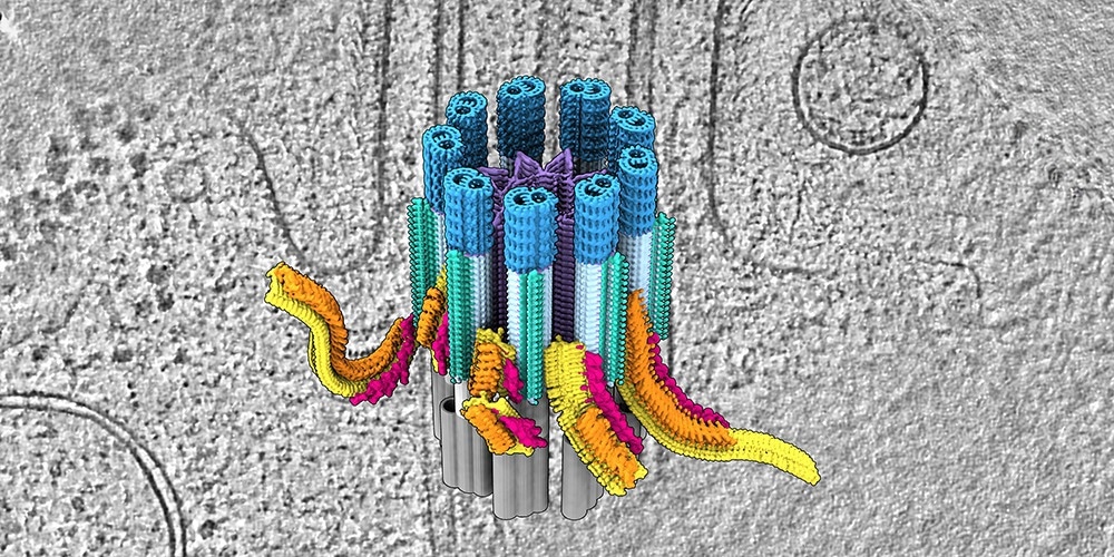
Cilia—small hair-like organelles—spread from cells and carry out many activities, which include signaling and motility. Scientists have discovered that cilia have a dedicated transport center at their base, where cargos and trains are gathered for transportation across the cilia. Because flaws in this cilia transport system can result in, for example, blindness or cystic kidneys, the findings reported in Science also offer new understandings of the molecular basis for different diseases.
 Cilia are highly complex transport hubs. Defects can lead to diseases e.g. cystic kidneys or blindness. Image credit: Biozentrum, University of Basel
Cilia do several activities for the cell: they aid cells in swimming, moving fluid, and sending messages to one another. They ensure sight, eliminate substances from the lungs, transport brain fluid, and smell and sound recognition. They are also important for the right development and arrangement of organs. When their functioning is interrupted, several different diseases can occur, which include kidney, heart, and lung diseases, infertility, or blindness.
The arrangement and working of cilia depend on large trains of proteins that transport vital cargos from and to the ciliary tip and the base, respectively. The traffic inside cilia can be paralyzed by even the tiniest mutations in individual components.
The study group headed by Prof. Ben Engel at the Biozentrum of the University of Basel, along with co-workers at the University of Geneva and the research institute Human Technopole in Milan, is now successful in investigating cilia in their usual environment. Their examination showed the native 3D structure of the ciliary base for the first time. The team found a busy transport center, with trains getting loaded and assembled to get ready for their transportation into the cilia.
Loading station for cilia transport
Cilia are strongly attached to the cell at their base.
Here is the start station for cilia transport. Trains are assembled here, loaded with cargo, and placed on the rails.”
Hugo van den Hoek, Study First Author, University of Basel
There are nine diverse rails in total within cilia, which are known as microtubules. Each comprises two tracks: each one for outbound and inbound trains. The trains pass on proteins, like signaling molecules, and building components to the cilia tip. The train is disassembled and unloaded at cilia’s destination station.
The team investigated the composition of the assembling trains in elaborate, which reveals the order in which the train components are assembled at the base of the cilia. Structures functioning as a selective barrier at the base are also imaged.
This regulates the entry of large trains until they are fully assembled and loaded with the cargo proteins required for the construction and maintenance of the cilia. From fluorescence microscopy, we also know the exact timetable of the trains. Trains leave the start station within nine seconds, and then the whole train assembly process starts again.”
Hugo van den Hoek, Study First Author, University of Basel
Organelles in 3D
The composition and structure of the ciliary base were resolved by researchers using two complementary imaging methods. The study teams of Ben Engel in Basel and Dr. Gaia Pigino in Milan conducted cryo-electron tomography, showing native cellular structures with delicate molecular detail.
Scientists headed by Dr. Virginie Hamel and Prof. Paul Guichard in Geneva included information from Expansion Microscopy, allowing several proteins to be mapped and localized onto the tomography structures.
This powerful combination of technologies has allowed us to reconstruct the first molecular model of the ciliary base and observe how it regulates the assembly and entry of these large protein trains.”
Paul Guichard, Professor, University of Geneva
“Understanding the transport system and its logistics in detail helps us understand how cilia are built and function, which may also provide new ideas for therapies to cilia diseases,” states Ben Engel. As a step forward, he and his co-workers want to investigate what happens at the tip of the cilia: structuring of this end station and organization of return transport.
Source:
Journal reference:
Hugo van den Hoek, et al. (2022) In situ architecture of the ciliary base reveals the stepwise assembly of IFT trains. Science. doi.org/10.1126/science.abm6704.