The protein SPOP, which also plays a role in endometrial, uterine, and other cancers, is the most mutated in prostate cancer. Despite its significance, it is still unclear how SPOP mutations cause cancer.
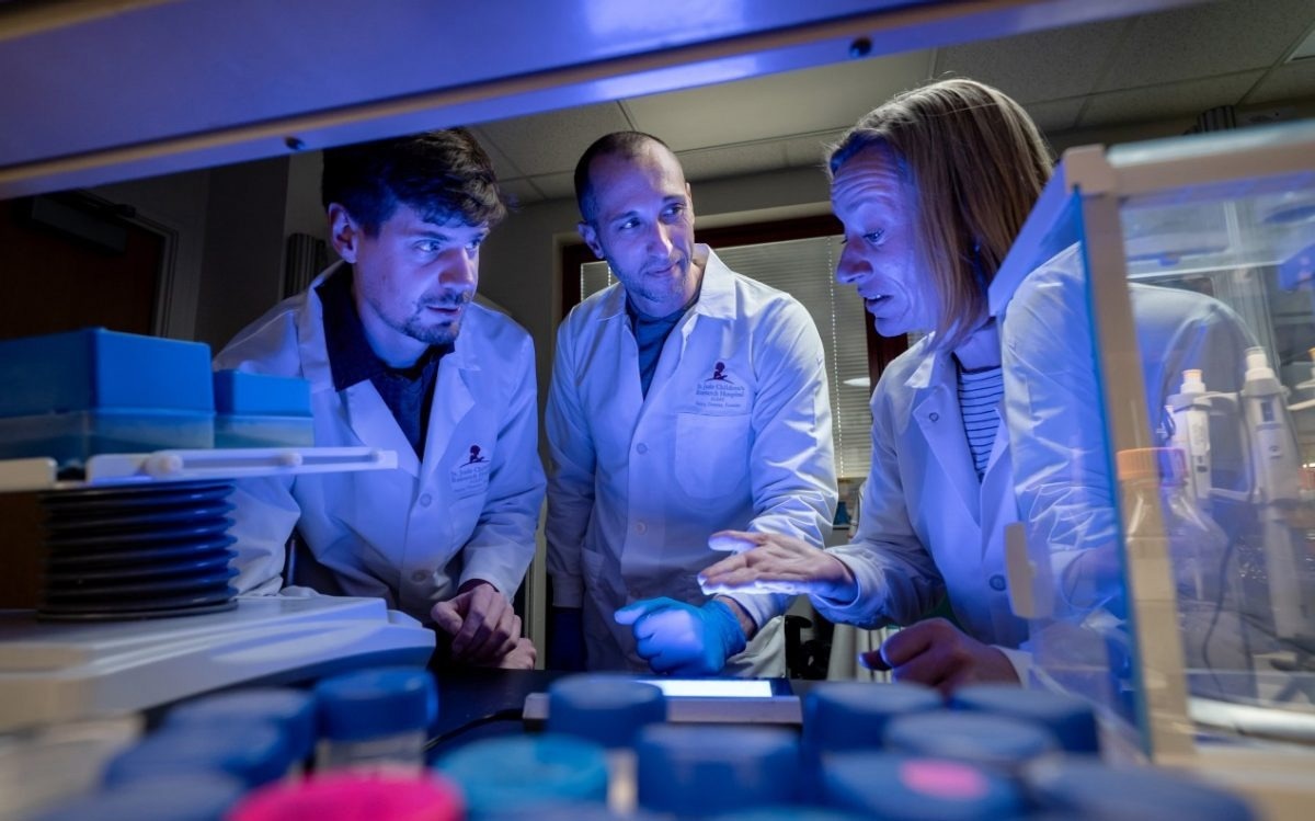
 L to R: Brian O’Flynn, PhD, Matthew Cuneo, PhD, and Tanja Mittag, PhD. Image Credit: St. Jude Children’s Research Hospital
Cryo-electron microscopy (cryo-EM) was used by researchers at St. Jude Children’s Research Hospital to record the first 3D structure of the entire SPOP assembly. The research, which was just published in Molecular Cell, identified previously unidentified SPOP interfaces that contain concentrations of cancer-causing mutations.
Controlling a cell’s level of particular proteins is one of SPOP’s typical roles. Since protein levels are altered and abnormal behaviors are triggered when SPOP is dysregulated as a result of mutations, it can have a dramatic impact on the cell. SPOP might not be the fire that ignites cancer, but it has the power to light the fuse if it is not properly controlled.
The prostate cancer-associated mutations are well understood. They are in the substrate-binding site and prevent SPOP from recognizing its substrates. But mutations found in patients with endometrial and other cancers were puzzling. The mutated sites did not seem to be important for SPOP function, at least looking at previous structures.”
Tanja Mittag, PhD, Study Corresponding Author and Member, Department of Structural Biology, St. Jude Children’s Research Hospital
Big picture understanding
The researchers were able to learn more about the underlying mechanisms that control the function of this protein in both its natural state and when it is mutated in cancer by determining the 3D structure of SPOP using cryo-EM.
The researchers were able to pinpoint significant interactions between previously unseen regions of the protein by observing how the SPOP oligomer comes together. These were precisely the regions that are mutated in endometrial cancer.
Once we had the 3D structure of SPOP, we could see that the pieces of the puzzle that had been missing were inherently important for understanding how SPOP functions in cancer. We were now able to see that what we initially thought to be regions of SPOP without functional importance are actually key to SPOP assembly and biology.”
Matthew Cuneo, PhD, Study Co-First Author, Department of Structural Biology, St. Jude Children’s Research Hospital
One mutation, dramatic effects
For instance, changes to the SPOP protein’s tendency to assemble and linger in cells were caused by changes to the MATH domain, which binds substrates. Additionally, the researchers noticed that a single mutation could have a significant impact on how the protein forms filaments.
The assembly of the proteins from a single filament to a double filament with intertwined MATH domains was completely altered by changing the interface that locks the MATH domain in place.
Through the use of cryo-EM, these extra protein interfaces in the filament were found, which furthered the understanding of how SPOP mutations cause cancer.
Taking a broader view to look at the full-length protein gave us a deeper understanding of how mutations affect SPOP. The scale of the change just from one point mutation is huge. It was unexpected to see that.”
Brian O’Flynn, PhD, Study Co-First Author and Postdoctoral Research Fellow, St. Jude Children’s Research Hospital
New questions
Drugs that target the MATH domain of SPOP have already been tested by researchers in the field. The mutant forms of SPOP in endometrial cancer could now be specifically targeted by drugs. As a result of the study, this could one day be possible to target SPOP differently depending on how it functions in the cell.
O’Flynn added, “In addition to the questions that this research answers, what is exciting is that for scientists asking about SPOP, the possibilities have really opened up.”
Source:
Journal reference:
Cuneo, M. J., et al. (2022). Higher-order SPOP assembly reveals a basis for cancer mutant dysregulation. Molecular Cell. doi.org/10.1016/j.molcel.2022.12.033