Researchers at St. Jude Children’s Research Hospital have solved the 3D structure of an important protein complex in cilia, which are signaling appendages found on cells. The structure was pictured at the highest resolution available at the time.
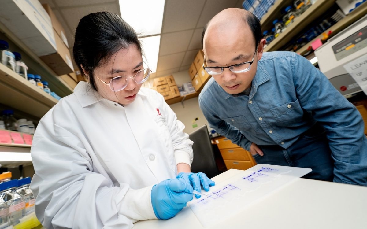
 First author Meiqin Jiang, PhD, and co-corresponding author Ji Sun, PhD, St. Jude Department of Structural Biology. Image Credit: St. Jude Children’s Research Hospital.
The findings will be used to explore diseases of the brain, kidney, skeleton, and eyes that are known to involve cilia but have been difficult to investigate. The results were published in the journal Cell Research.
Cilia are hair-like projections observed on almost all mammalian cells that regulate a variety of signaling processes. The cilium is a thin tube that lacks the machinery needed to synthesize proteins required for cell signaling. As a result, signaling molecules within cilia must be transported from other parts of the cell. IFT-A and IFT-B are intraflagellar transport complexes.
Researchers have been trying to figure out the structure of the IFT-A and -B complexes for over a decade. The structure of IFT-A was ascertained by St. Jude researchers to an overall resolution of around 3–4 Angstroms, allowing us to visualize these complexes in near-atomic detail.
Now that we have the high-resolution structure of this ciliary complex, we can map mutations known to cause diseases and then design clinical interventions. We can tell at the atomic level tell how these trains assemble with each other to form very elegant structures in the cilia. We can also use that knowledge to understand how disease mutations disrupt that structure.”
Ji Sun PhD, Co-Corresponding Author, Department of Structural Biology, St. Jude Children’s Research Hospital
Their research illustrates new details that were never obvious enough in previous attempts. They discovered previously unknown zinc-binding sites in IFT-A, for example. Zinc-binding sites are essential for zinc fingers, a type of protein domain. Zinc fingers are required for certain protein–protein interactions, explaining some previously unknown connections within the train complex.
It was pretty exciting to see the zinc fingers because no one had seen or even predicted zinc-binding sites in IFT-A. Our study was able to reveal that with confidence. We could say, ‘Hey, there’s zinc, and it is important for protein-protein interaction, which might also facilitate train assembly.’ We never would have figured that out without our high-resolution structural information.”
Ji Sun PhD, Co-Corresponding Author, Department of Structural Biology, St. Jude Children’s Research Hospital
A molecular ticket to ride
The train, while essential, is only one part of a story. The structure of IFT-A in complex with the protein Tubby-related protein 3 (TULP3) was solved by the scientists.
To ride a train, you need a ticket. TULP3 is a ticket to get on the train of IFT-A. TULP3 can then recognize different acceptable cargo to be transported on the train. So, when you have molecular cargo that can hold onto this ticket, it can ride the train. If you disrupt this TULP3 interaction, then you can no longer transport certain cargos, because they lack a valid molecular ticket.”
Ji Sun PhD, Co-Corresponding Author, Department of Structural Biology, St. Jude Children’s Research Hospital
The discovery of the complex 3D structure of TULP3 and IFT-A is a significant achievement that will provide insight into how signaling molecules travel to and from cilia, and how disruption in the interface between the two can cause disease.
Understanding diseases of cilia at a high resolution
Cilia are vital organelles in several species. Researchers believe that cilia are essential because of their high conservation across species. Numerous ciliary protein mutations are linked to diseases of various tissues. However, without a structure, it is difficult to explain how protein changes cause disease.
As a result, scientists have made numerous attempts over the last decade to solve the structure of ciliary components. Using a technique known as single-particle electron cryo-microscopy (cryo-EM), the St. Jude team was able to create a high-resolution structure of IFT-A.
Sun says, “The field has been waiting a long time to see these complexes. IFT-A is a complex of six proteins. We know that IFT-A mutations affect skeletal development, especially the ribs, but also structures like the retina. There are many developmental diseases caused by mutations in this complex.”
The high-resolution structures of IFT-A and TULP3 in a complex can now serve as the foundation for research into several developmental diseases involving cilia and aid in the development of novel strategies to mitigate or cure them.
Source:
Journal reference:
Jiang, M., et al. (2023) Human IFT-A complex structures provide molecular insights into ciliary transport. Cell Research. doi.org/10.1038/s41422-023-00778-3.