Reviewed by Danielle Ellis, B.Sc.Jan 3 2024
Researchers at St. Jude Children’s Research Hospital captured images of cytonemes, which are cellular projections that are involved in long-distance signal transmission during development.
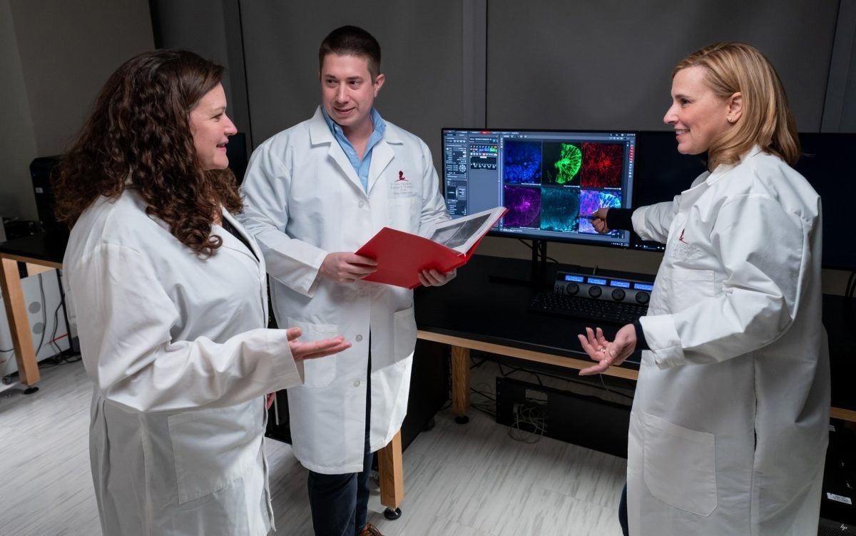
 (L to R) Second author Miriam Dillard, first author Eric Hall, PhD, and corresponding author, Stacey Ogden, PhD, St. Jude Department of Cell and Molecular Biology. Image Credit: St. Jude Children’s Research Hospital
Scientists at St. Jude Children’s Research Hospital have discovered the significance of cytonemes- long, thin projections on cells that resemble hairs - during the development of the brain.
Although cytomemes are difficult to see under a microscope in the tissues of developing vertebrates, they link cells that communicate over great distances. The researchers have made the first visual representation of the transport of signaling molecules by cytonemes during the development of the mammalian nervous system. The research results were released in Cell.
We showed cytonemes are a direct express route for signal transport. Cells need to communicate with each other during development and tissue homeostasis and be able to reach more than just their neighbors. We’ve identified one way that signals are loaded into cytonemes for transport to responding cell populations and demonstrated that tissue patterning does not happen properly when this mode of signal dispersion is compromised.”
Stacey Ogden PhD, Corresponding Author, Department of Cell and Molecular Biology, St. Jude Children’s Research Hospital
Express Signal Delivery Sets Up the Nervous System For Success
Mammalian cytonemes in the developing nervous system were first seen by researchers in Ogden’s lab using a combination of advanced microscopy methods and well-executed sample preparations.
For a long time, visualizing these structures in developing mammalian tissue has been challenging. But we’ve finally found a way.”
Stacey K. Ogden, Corresponding Author, Department of Cell and Molecular Biology, St. Jude Children’s Research Hospital
With the help of the novel techniques, the researchers were able to take pictures of how cytonemes function as a kind of “express” system that can bypass nearby cells in order to send signals directly to farther-off ones, like an express train that only stops at major hubs.
The notochord is one of the main stations; it generates a signal that is essential for structuring the growing spinal cord. Images of the transport process occurring in the cytonemes that originate from the notochord were obtained by Ogden’s team.
In mouse models, the researchers disrupted neural development and caused significant neurological defects by preventing signaling proteins from entering cytonemes.
This is the first demonstration of these cytoneme-based transport processes occurring during the development of a complex mammalian tissue such as the neural tube. Then we showed that when we reduce cytoneme numbers or decrease the ability of cells to load signaling proteins into these structures, we get developmental defects.”
Stacey K. Ogden, Corresponding Author, Department of Cell and Molecular Biology, St. Jude Children’s Research Hospital
Cytoneme Transport Helps Establish the Gradient
The process of mammalian development is meticulously regulated and requires coordination in order for all tissues and organs to form properly. A mechanism by which cells determine when to take on a particular destiny is by reacting to specific thresholds of signaling proteins known as morphogens.
Across a signaling gradient, cells will react to these signals differently, assuming distinct characteristics in response to high and low concentrations of a given morphogen. Development requires a good gradient; a poor gradient can lead to catastrophe.
The process by which these morphogen patterns are generated in various fields of cell organization remains a mystery despite their significance. Some of these gradients can be explained by simple diffusion, but not all of them. The evidence supporting the critical role of cytoneme signaling during morphogen patterning is provided by the neurological deficits that resulted from the scientists’ blocking of signals entering cytonemes.
Ogden says, “These deficits are really the first direct evidence that cytoneme-based signaling plays a key role during neural tube patterning.”
Transporting “Sonic Hedgehog” Through Cytonemes
One method by which a gradient of the sonic hedgehog morphogen forms in the neural tube was elucidated by the St. Jude group. Although the path taken by Sonic Hedgehog to reach its target cells has not always been clear, it was previously recognized as a crucial signaling molecule in the development of the brain.
According to the study, cytonemes are an essential component of the mechanism that generates the sonic hedgehog signaling gradient. Accordingly, Sonic Hedgehog’s signaling function was hampered when it was unable to be loaded into Cytonemes.
Odgen continues, “In the morphogen signaling research field, we’ve always wanted to know how a signal gets from one population of cells to spread across a receiving cell population to make a gradient. It’s really exciting to show that cells that are producing morphogen signals are playing an active role in getting them to where they need to go through cytonemes. The signaling cell is not only making the morphogen, but it’s also helping to physically deliver the signal.”
The extent to which cytoneme express transport of signaling proteins is being developed is still unknown.
Ogden says, “Here, we’ve used sonic hedgehog as a model. But we also have evidence that these structures may be important for transporting other signals that are crucial during neural tube development. Now that we’ve developed a system to visualize these cytonemes, we can begin to uncover the true breadth of their function.”
Source:
Journal reference:
Hall, T. E., et al. (2023). Cytoneme signaling provides essential contributions to mammalian tissue patterning. Cell. doi/S0092-8674(23)01333-8#%20